
 United Kingdom vs Turkey
 United States vs Turkey
 Germany vs Turkey
 France vs Turkey
 Spain vs Turkey
 Sweden vs Turkey
 Denmark vs Turkey
 Finland vs Turkey
 Ireland vs Turkey
 Netherlands vs Turkey
 Norway vs Turkey
 Australia vs Turkey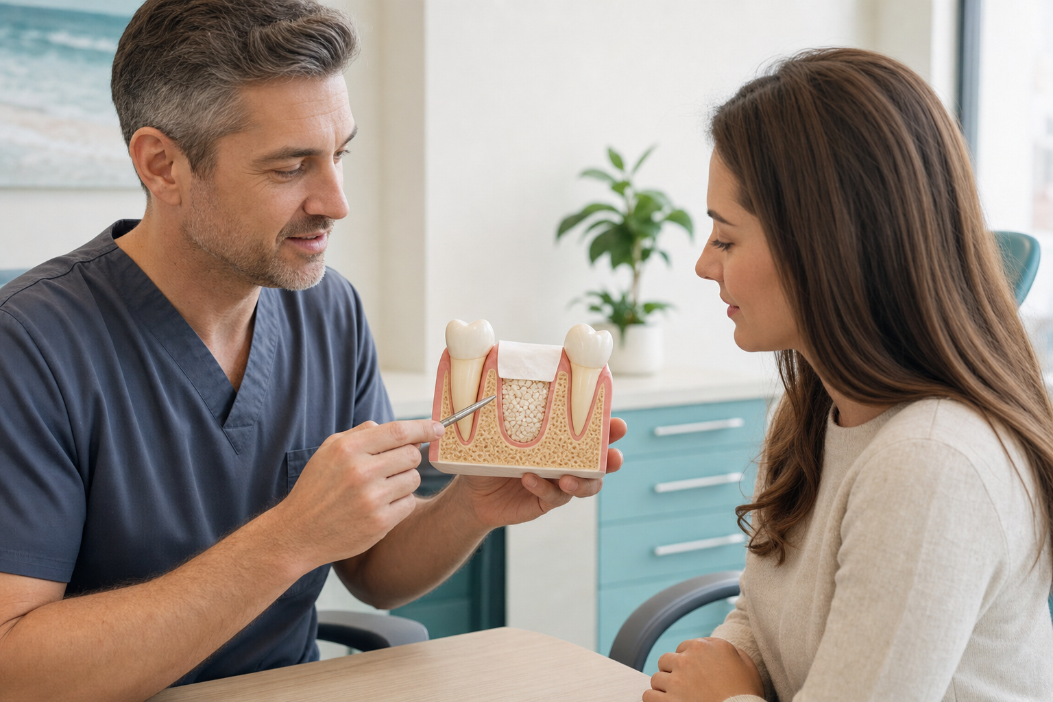
Clinical review required: This draft is educational. Socket management must be selected after diagnosis of the tooth, socket walls, infection, soft tissue and future restorative plan.
What is socket preservation?
Socket preservation, also called alveolar ridge preservation, is performed at or shortly after tooth extraction to reduce the normal loss of ridge width and height. After a tooth is removed, the socket fills with clot and new bone, but its outer contours remodel, often most on the facial side. A clinician may place graft material and sometimes a membrane or soft-tissue seal to support the space. The procedure reduces average dimensional change; it does not freeze the ridge or reproduce the original tooth-supporting bone exactly.
Why the ridge changes after extraction
The thin bundle bone lining the socket depends partly on the periodontal ligament and remodels after tooth loss. Facial plates can be very thin or missing, especially around front teeth and infected roots. Soft tissue follows the underlying contour. The amount of change varies with anatomy, trauma, disease, tooth type and patient factors. Immediate implant placement alone does not stop this biology.
When preservation may be useful
It is commonly considered when an implant is planned later, when ridge contour supports an aesthetic bridge or denture, or when treatment timing is uncertain but future options should be protected. It may reduce the need or extent of later augmentation. Benefit is greater when dimensional loss would compromise a known restorative plan. Routine grafting of every socket is not automatically necessary.
When it may not be needed
A socket may heal naturally when no replacement is planned, adequate bone will remain, immediate treatment is appropriate or grafting would not change the restorative decision. Acute infection, major wall loss or soft-tissue deficiency may require a different regenerative approach rather than simple socket filling. Cost, additional material and potential complications should be justified by expected benefit.
Diagnosis before extraction
The clinician evaluates restorability first; preservation should not encourage unnecessary extraction. Examination and imaging identify root anatomy, infection, facial-plate condition, periodontal attachment, adjacent structures and future crown position. CBCT is selected when three-dimensional information will affect treatment, not automatically for every socket. The consent process should distinguish extraction, socket preservation and later implant placement as separate decisions.
Atraumatic extraction
Preserving socket walls begins with controlled removal. Sectioning multi-rooted teeth, using periotomes or fine instruments and avoiding uncontrolled leverage can reduce fracture. Granulation tissue and pathology are removed while vital walls are protected. “Atraumatic” does not mean no trauma; it describes deliberate techniques intended to limit avoidable damage.
Graft materials
Autograft, allograft, xenograft and synthetic alloplasts may be used alone or mixed. Slowly resorbing mineral can support contour but leave particles; faster-remodelling materials may be replaced sooner but lose volume. Collagen plugs and matrices can stabilise clot or particles. The exact material, biological origin, processing and expected remodelling should be disclosed. A product is a scaffold, not a guarantee of vital bone.
Membranes and socket seals
A resorbable membrane may cover a deficient wall or contain graft. Dense non-resorbable barriers can sometimes remain exposed intentionally but require removal. Free gingival grafts, connective-tissue grafts, collagen matrices or carefully designed flaps can seal the entrance. Primary closure is not always required, and advancing a flap may alter the gum line or vestibule. Technique should match the aesthetic and implant plan.
Preservation with an intact socket
When walls are intact, particles are placed gently without excessive compression and the entrance is stabilised. Overpacking can impair vascular ingrowth or push material beyond boundaries. The graft should support contour, not create an oversized ridge. Soft tissue grows across the top while bone develops from socket walls.
Sockets with missing walls
A large facial dehiscence is closer to ridge augmentation than simple preservation. A membrane, tenting, additional graft and soft-tissue management may be needed. Infection control and closure become more important. Patients should understand that severe damage can still require secondary augmentation even after careful treatment.
Infected sockets
Chronic periapical or periodontal infection does not always prohibit grafting if the source is removed and the site can be thoroughly debrided. Spreading infection, uncontrolled drainage or inability to clean the defect may favour delayed regeneration. Antibiotics do not replace debridement. The decision is based on clinical findings, systemic risk and the planned material.
Socket preservation versus immediate implant
An immediate implant can shorten treatment but requires correct three-dimensional placement and primary stability. It does not fill the socket or prevent facial remodelling automatically. Preservation followed by delayed implantation can provide time for infection and soft tissue to resolve, but adds a later operation. Neither pathway is universally superior. A 2019 review found heterogeneous comparisons, reinforcing patient-specific selection.
Does preservation prevent later grafting?
It can reduce the likelihood or extent of augmentation but cannot promise avoidance. Thin facial plates, large defects, graft resorption and implant-position requirements may still leave a deficiency. The correct outcome is enough healthy, maintainable tissue for the restoration, not merely a socket that looks radiopaque.
Healing and timing
Soft tissue closes over weeks while mineralisation continues for months. Implant placement is commonly planned after several months, adjusted for material, defect and medical factors. Early placement may use developing bone; later placement allows maturation but also continued remodelling. Clinical and radiographic reassessment should determine timing rather than a fixed package calendar.
How healing is assessed
The clinician checks closure, inflammation and contour. At implant surgery, tactile preparation and direct observation provide information about tissue quality. Histology is rarely needed clinically. CBCT shows dimensions and mineral density but cannot by itself distinguish every residual particle from vital bone. Implant stability must come from appropriate bone, not a favourable-looking scan alone.
Pain and recovery
Discomfort usually reflects the extraction and any flap or donor procedure. Mild swelling, oozing and bruising are expected. A collagen plug or small particles may be visible early. Increasing pain after improvement, fever, pus, bad taste or progressive swelling needs assessment. Not every loose surface particle means the entire graft has failed.
Possible complications
- Bleeding, dry socket, infection or delayed closure.
- Membrane exposure and loss of graft particles.
- Insufficient width or height despite preservation.
- Soft-tissue recession, scar or altered vestibular depth.
- Material sensitivity or donor-site morbidity, depending on graft.
- Damage to adjacent structures during extraction.
- Need for regrafting or a revised implant plan.
Dry socket and preservation
Alveolar osteitis causes increasing deep pain several days after extraction when the clot breaks down. Grafting does not eliminate the risk. Smoking, previous dry socket and traumatic removal matter. Treatment focuses on assessment, gentle irrigation, analgesia and local care; antibiotics are not the routine cure unless infection is present.
Medical and lifestyle factors
Smoking and nicotine impair blood supply and increase wound risk. Poorly controlled diabetes, immune suppression, periodontal disease and inadequate hygiene require management. Antiresorptive or antiangiogenic medicines and previous radiotherapy need individual extraction-risk assessment. Patients should disclose all medicines and not stop them independently.
Evidence and realistic expectations
Systematic reviews generally find less average horizontal and vertical loss with preservation than spontaneous healing. Results vary by tooth, wall anatomy, technique, material and measurement. The procedure does not eliminate remodelling, and evidence that one biomaterial is universally best is limited. Implant survival can be high after either pathway; the clinically relevant advantage is often a more favourable implant site or less later surgery.
PRF in extraction sockets
Platelet-rich fibrin can be placed as clots or membranes and may support early soft-tissue healing and reduce dimensional loss compared with spontaneous healing. A 2026 meta-analysis reported favourable changes at several time points, but protocols and certainty varied. PRF alone may be reasonable for selected sockets; it should not be represented as a structural substitute for graft in every large defect.
Aftercare
Bite on gauze as directed, protect the clot and eat soft food. Avoid smoking, vaping, vigorous rinsing, straws and strenuous activity initially. Begin cleaning and prescribed rinses at the advised time. Do not pick at membranes or particles. A temporary denture must be relieved so it does not press on the socket.
Front teeth and aesthetic risk
In the smile zone, preservation may support facial contour but cannot guarantee the final gum margin or papilla. Thin facial bone, a high smile line, recession, infection and a facially positioned root increase risk. Soft-tissue grafting, provisional restoration design and implant timing may be as important as the socket filler. A bulky graft cannot correct an implant placed too far facially. Photographs, diagnostic tooth position and adjacent gum levels should therefore form part of planning.
Molar sockets
Multi-rooted molars leave wide sockets with interradicular bone and differing wall thickness. The surgeon may preserve the entire site, graft selected compartments or plan a future implant around the septal bone. Maxillary molars can lie close to the sinus, while mandibular molars approach the inferior alveolar nerve. Material must not be forced into protected anatomy. A wide socket also takes longer to mineralise than its soft-tissue appearance suggests.
Temporary teeth during healing
A removable temporary tooth, bonded bridge or existing denture may be used, but it must not compress the graft or wound. Pressure can open the seal, displace particles and flatten the desired contour. A provisional appliance may need relining or repeated adjustment as swelling resolves. The patient should understand whether the temporary is cosmetic only and must avoid chewing on it until the team confirms safe function.
Long-term value and cost
Preservation adds material, clinical time and sometimes membrane removal, yet it may reduce a later augmentation. Economic value depends on whether the retained dimensions change the final treatment. Patients should compare the complete pathway: extraction, graft, temporary restoration, imaging, implant, possible additional graft and final crown. A lower initial package price is not necessarily cheaper if it excludes review or complication care. Conversely, routine grafting without a future restorative benefit adds cost without clear purpose.
Treatment abroad
Request the reason for preservation, socket-wall assessment, exact graft and membrane, closure method and intended implant timing. Obtain product traceability and operative notes. Stay for early review. Clarify who treats dry socket, infection, exposure or insufficient bone after return. Preservation is not proof that an implant can be placed without reassessment.
Questions to ask
- Is the tooth definitely non-restorable?
- Which socket walls are intact?
- How will preservation change my future treatment?
- What material and origin are proposed?
- Will a membrane or soft-tissue seal be used?
- Could the socket heal naturally?
- When will implant suitability be reassessed?
- What happens if particles or membrane expose?
Frequently asked questions
Does socket preservation stop all shrinkage?
No. It reduces average dimensional change but cannot prevent normal remodelling completely.
Is it the same as immediate implantation?
No. Preservation manages the socket; an immediate implant is a device placed at extraction and may still require grafting.
Will graft particles fall out?
A few superficial particles can appear, but persistent loss or wound opening needs review.
Does a graft guarantee enough bone?
No. Healing and the later implant position determine whether additional augmentation is required.
Sources and clinical review references
- Effects of alveolar ridge-preservation interventions.
- Biomaterials for ridge preservation: network meta-analysis.
- Implants in preserved versus fresh extraction sockets.
- PRF alone for alveolar ridge preservation.
Editorial review note: Evidence reviewed 22 July 2026. Named clinician review is required before indexation.