
 United Kingdom vs Turkey
 United States vs Turkey
 Germany vs Turkey
 France vs Turkey
 Spain vs Turkey
 Sweden vs Turkey
 Denmark vs Turkey
 Finland vs Turkey
 Ireland vs Turkey
 Netherlands vs Turkey
 Norway vs Turkey
 Australia vs Turkey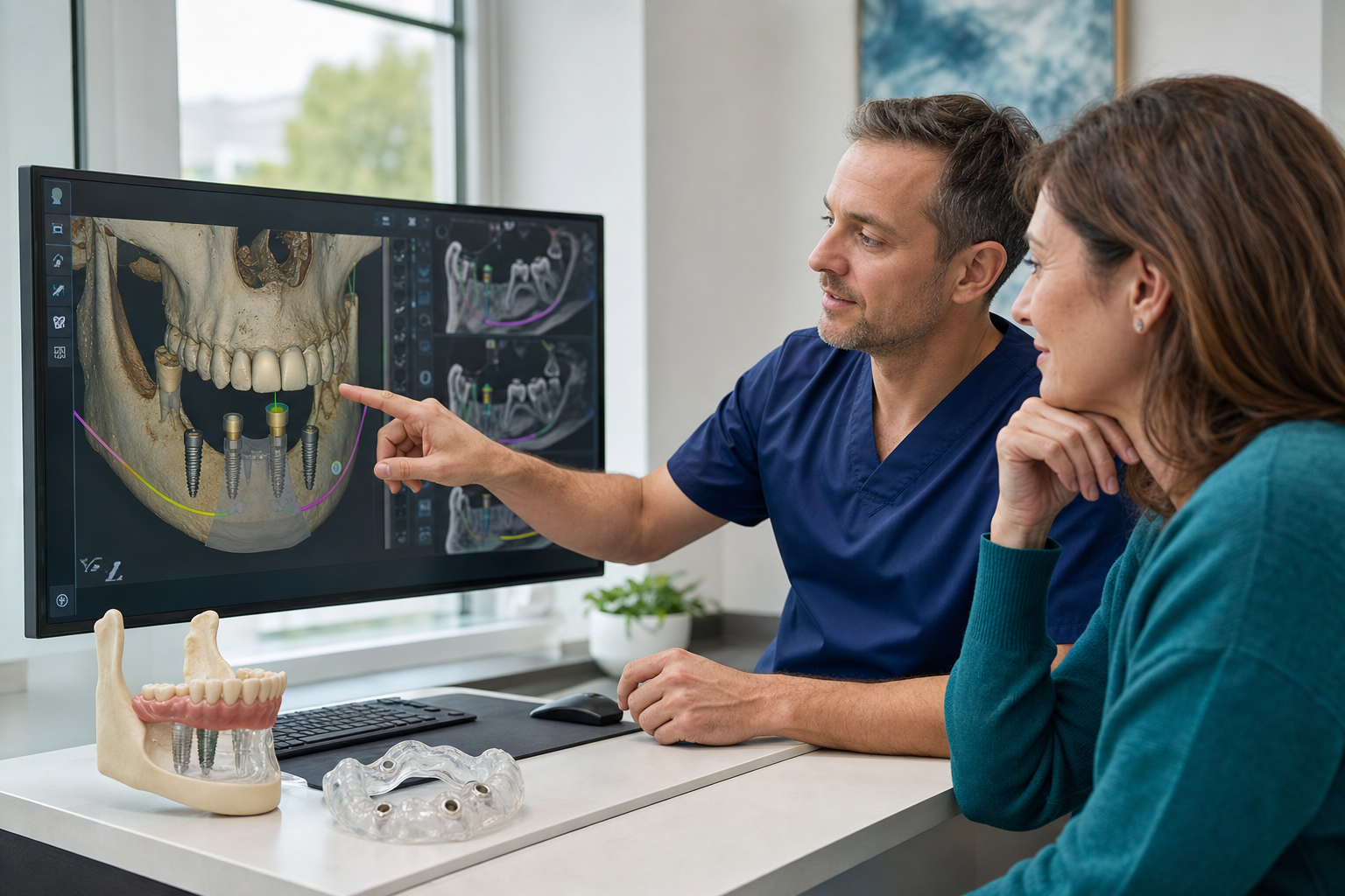
Definition: Computer-guided implant surgery uses digital imaging and planning software to design implant positions and transfer them with a static guide, dynamic navigation or robotic assistance. It improves planning precision but does not eliminate deviation or surgical judgement.
What is computer-guided implant surgery?
The workflow combines three-dimensional anatomy with a digital representation of future teeth. The clinician plans implant position, diameter, length and angulation relative to bone, nerves, sinuses and the prosthesis. The plan is transferred during surgery using a manufactured guide or real-time tracking system.
Why start with the final tooth?
Prosthetically driven planning places the implant under the intended crown or bridge, aiming for suitable screw access, emergence and cleanability. Bone anatomy may require compromise, grafting or a different prosthesis. A technically safe implant that cannot support a maintainable tooth is not a complete success.
The digital data sources
CBCT provides three-dimensional information about mineralised structures. An intraoral or model scan records tooth and soft-tissue surfaces with greater surface detail. A diagnostic setup defines the planned teeth. Software merges these datasets. Each source has error, and inaccurate merging propagates into the surgical plan.
CBCT is not automatic
Radiation should be justified by the clinical question and field of view. Artefact from metal can obscure boundaries. The clinician must review the entire acquired volume, not only the implant slice. A CBCT alone does not capture mobile soft tissue or guarantee nerve visibility.
Static computer-assisted surgery
A static guide contains sleeves or channels that constrain drills and sometimes implant insertion. Guides may be tooth-, mucosa- or bone-supported. Tooth support is often stable when sufficient teeth remain. Mucosa-supported full-arch guides require fixation and can move on compressible tissue.
Dynamic navigation
Dynamic systems track the patient and drill in real time, displaying deviation from the plan. They allow intraoperative angle changes and avoid a bulky guide. Accurate registration, calibration and uninterrupted tracking are essential. The surgeon must coordinate screen information with the operative field.
Robotic assistance
Robotic systems can provide haptic or mechanical guidance along the planned trajectory. Evidence is evolving, and availability and training vary. Higher technological complexity does not automatically improve survival or make a poor restorative plan appropriate.
Fully guided, pilot guided and partially guided
Fully guided protocols control the full drill sequence and implant insertion. Pilot guides control only the initial osteotomy; later steps are freehand. Partially guided workflows use the guide for some drills. Patients should know which is proposed because “guided” does not describe a single level of control.
Data acquisition workflow
- Clinical examination and disease control.
- CBCT using appropriate positioning and field.
- Intraoral or model scan.
- Diagnostic tooth setup.
- Dataset registration and verification.
- Implant planning with safety margins.
- Guide design or navigation registration.
- Surgical verification and postoperative assessment.
Dataset matching
Software aligns surface scans with CBCT anatomy using teeth, markers or a dual-scan denture protocol. The clinician checks several regions rather than accepting automatic alignment. A visually plausible overlay can still be wrong. Full-arch edentulous cases are harder because stable tooth landmarks are absent.
Planning safety margins
Guided placement has coronal, apical, depth and angular deviations. Therefore implants are not planned touching nerves, roots or cortical boundaries. The required margin depends on anatomy, system evidence and clinician judgement. Planning to the theoretical edge removes tolerance for accumulated error.
Guide support and fixation
A tooth-supported guide is checked for complete seating. Mucosa-supported guides can compress or rock and often use fixation pins. Bone-supported guides require flap exposure. Inspection windows, reference stops and a repeatable seating protocol help detect error.
Sleeves, keys and drill tolerance
Clearance between drill, key and sleeve permits rotation but contributes to deviation. Long sleeves may improve guidance but reduce irrigation or access. Limited mouth opening can prevent insertion of long drills. The virtual plan must be physically feasible in the patient's mouth.
Flapless surgery
Computer guidance may support flapless placement through a small tissue punch. Potential benefits include less surgical exposure and early discomfort. The surgeon cannot directly see bone contours, defects or tissue thickness. A flap should be raised when visibility, grafting or safety requires it.
Guided bone reduction
Full-arch systems may guide alveoloplasty as well as implants. Bone reduction establishes restorative space but is irreversible. The guide must seat at a verified reference before bone is removed. Patients should understand the planned reduction and its relationship to the prosthetic transition line.
Immediate provisional workflow
Accurate planned placement can support prefabricated provisional teeth. Actual positions are verified, and the prosthesis may need conversion or adjustment. A guide does not guarantee adequate primary stability or passive fit. A backup temporary remains necessary.
Accuracy evidence
Systematic reviews generally find static and dynamic computer-assisted systems more accurate than freehand placement. Mean deviations remain clinically meaningful, especially at the apex. Accuracy does not necessarily translate into higher implant survival or less marginal bone loss; those outcomes have been comparable in some reviews.
Fully edentulous accuracy
Edentulous cases lack tooth support and can show greater guide movement and scan-registration difficulty. A 2024 review reported larger apical than cervical deviations and a tendency toward superficial placement in included full-arch studies. Fixation and verification are therefore critical.
Benefits
- Visualises implant, anatomy and prosthesis in one plan.
- Can improve positional accuracy and prosthetic access.
- May support flapless or minimally invasive surgery in selected cases.
- Facilitates digital provisional and team communication.
- Useful for limited space and multiple implants when properly verified.
Limitations and risks
Errors can arise from imaging, segmentation, scanning, merging, guide manufacture, guide seating, drilling and patient movement. Guides can fracture, interfere with irrigation or limit visibility. Dynamic navigation can lose tracking. Technology adds cost and a learning curve. Conversion to freehand surgery must remain possible.
Does guided surgery prevent nerve injury?
It can improve planning and trajectory control but cannot guarantee prevention. Nerve identification, safety margins, guide stability and intraoperative judgement remain essential. Persistent numbness after surgery requires urgent assessment regardless of the method used.
Postoperative verification
The clinician records actual implant position and component seating. Additional imaging is justified only when it answers a clinical question; routine postoperative CBCT adds radiation. Deviations affecting the prosthesis may require angled components, redesign or, rarely, implant revision.
Segmentation and anatomical interpretation
Planning software converts scan greyscale into surfaces, but bone and nerve boundaries are not always automatically reliable. Thin cortical plates may disappear with threshold changes. The clinician reviews multiplanar slices and corrects segmentation where needed. A smooth three-dimensional rendering can conceal uncertainty present in the source images.
Metal artefact and missing data
Crowns, posts and existing implants can create streaks that disrupt surface matching or obscure roots. Repeat scanning is not automatically the solution because it adds radiation and may reproduce artefact. Alternative landmarks, radiographic markers, conventional impressions or a modified guide design may be required.
Navigation registration and calibration
Dynamic navigation links the patient's anatomy to the virtual plan using a reference marker and calibrated handpiece. Registration error affects every osteotomy. The system is checked against known landmarks before drilling and recalibrated if the marker moves or instruments change. Real-time display is only as accurate as this relationship.
Learning curve and human factors
Static guidance changes tactile feedback and irrigation; dynamic navigation requires hand-eye coordination while monitoring a screen. Robotic systems require workflow-specific training. Experience with freehand anatomy remains important for recognising when the technology is wrong. Clinics should maintain simulation, calibration and emergency-conversion competence.
Digital planning for immediate loading
The virtual implant plan may be used to prefabricate abutments or a provisional bridge. This creates an accuracy chain from imaging to scan merge, guide or navigation, implant carrier and prosthesis. Each tolerance accumulates. Intraoperative verification and a convertible temporary reduce dependence on a perfect digital transfer.
Stacked and anchored guide systems
Full-arch workflows can use a fixed base supporting sequential bone-reduction, implant and prosthetic guides. The base must be placed at the correct vertical and facial reference. If it is wrong, later guides can fit perfectly to the wrong position. Reference pin verification and visual checks are essential at every stage.
Data ownership and export
Patients should receive implant records and clinically useful images. Ask whether DICOM, scan and plan files can be exported in standard formats or are locked to proprietary software. Archived data can help remake a guide or prosthesis, but a future clinician must still verify that anatomy and teeth have not changed.
Cost-benefit discussion
Computer guidance adds imaging, planning, manufacturing and equipment costs. Its value is greater when anatomy is constrained, several implants must coordinate with a prosthesis or flapless access is justified. A simple site with generous anatomy may not gain the same benefit. The clinic should explain the clinical reason, not only the technology brand.
Accuracy reporting that patients can understand
Accuracy studies commonly report deviation at the implant entry point, deviation at the apex, angular deviation and depth error. These measurements describe different aspects of placement and should not be reduced to a claim that a system is simply “precise.” A small angular difference can create a larger apical displacement as implant length increases, while a modest coronal shift may matter greatly beside a neighbouring tooth.
Published averages also contain a range. A clinic should plan for plausible deviation rather than placing the virtual implant directly against a nerve, sinus boundary or adjacent root. Guidance can improve transfer of a plan, but safety still comes from margins, verification and the willingness to stop or convert when the guide does not behave as expected.
Questions for a digital implant clinic
- Is the surgery static, dynamic, robotic or only pilot guided?
- How are CBCT and surface scans registered and checked?
- What support and fixation does the guide use?
- What safety margins are planned?
- What is the backup if the guide does not seat?
- Does the provisional depend on exact guided placement?
Treatment in Turkey
Ask who performs segmentation and planning and whether the surgeon approves the final plan. Request screenshots showing the prosthesis and anatomy, not marketing animations alone. Keep the guide plan, implant coordinates, component records and postoperative images for continuity of care.
Frequently asked questions
Is computer-guided surgery more accurate?
On average, guided methods can improve positional accuracy, but deviations remain and clinical outcomes also depend on biology and maintenance.
Does it mean no incision?
No. Flapless surgery is possible in selected cases, but a flap may be required for visibility, grafting or safety.
Can the computer place the implant?
Software plans; the clinician executes and verifies. Some robotic systems assist movement, but professional judgement remains responsible.
Sources and clinical review references
- Yogui FC, et al. Computer-guided versus freehand implant placement. Int J Oral Maxillofac Surg. 2021.
- Khan M, et al. Accuracy of robotic, static and dynamic implant guidance. J Prosthet Dent. 2024.
- Pellegrino G, et al. Dynamic navigation in implant dentistry. Int J Oral Maxillofac Implants. 2021.
- Azevedo M, et al. Accuracy of fully guided surgery in edentulous patients. J Clin Med. 2024.
Content status: editorial draft for review by a licensed dentist before publication. Last evidence check: 22 July 2026.