
 United Kingdom vs Turkey
 United States vs Turkey
 Germany vs Turkey
 France vs Turkey
 Spain vs Turkey
 Sweden vs Turkey
 Denmark vs Turkey
 Finland vs Turkey
 Ireland vs Turkey
 Netherlands vs Turkey
 Norway vs Turkey
 Australia vs Turkey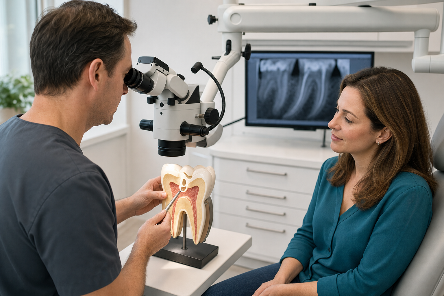
Clinical scope: A dental operating microscope provides coaxial illumination and variable magnification during endodontic diagnosis and treatment. It can improve visual access and technical control, especially in complex cases. It is an adjunct to training, anatomy, disinfection and restoration, not a treatment type or guarantee by itself.
What is microscope root canal treatment?
The phrase describes root canal treatment performed with a dental operating microscope. The biological goals remain diagnosis, canal detection, disinfection, filling and coronal sealing. The microscope lets the clinician view the access cavity and selected canal areas at magnification while maintaining an ergonomic working position.
How is it different from dental loupes?
Loupes provide fixed lower magnification and move with the clinician’s head. A microscope offers adjustable magnification, strong coaxial light and a stabilised optical field. Both can improve visualisation over unaided vision. A microscope does not see through tooth or around every curve; radiographs, tactile feedback and anatomy remain necessary.
Low, medium and high magnification
Low magnification provides orientation and a broader field during access. Medium magnification supports chamber-floor inspection and routine ultrasonic work. High magnification can examine a fine crack, canal opening or repair margin but has a narrow field and shallow depth of focus. The clinician changes levels rather than performing the entire procedure at maximum power. More enlargement is not automatically more accurate: loss of orientation or movement outside the tiny visual field can increase risk. Illumination, mirror position, patient stability and operator training determine whether a chosen level is useful.
Why illumination matters
Coaxial light travels close to the viewing axis and reduces shadows inside the access cavity. This can reveal colour changes, developmental grooves, canal orifices and fine cracks. Visibility still depends on a clean dry field. Blood, restorative debris and a narrow access can obscure anatomy even at high magnification.
When is a microscope especially useful?
Common indications include calcified canals, missed anatomy, retreatment, post and separated-instrument removal, perforation repair, resorption and endodontic microsurgery. It also supports routine access refinement and documentation. The greater the technical complexity and consequence of unnecessary dentine removal, the more useful controlled magnification may be.
Does every root canal need a microscope?
Many straightforward cases can be treated successfully with suitable illumination and magnification by trained clinicians. A microscope may enhance routine work, but evidence does not establish that every case without one is substandard or will fail. Referral decisions consider anatomy, operator skill, previous treatment and expected technical difficulty.
Finding hidden canals
Magnification helps inspect the chamber floor and trace grooves conservatively. Maxillary molars may contain a second mesiobuccal canal; mandibular teeth can have additional anatomy. Recent evidence associates missed canals with apical disease, though study certainty has limitations. Finding an orifice is only the start; the canal must be negotiated and disinfected safely.
Calcified canals
Calcification can narrow or obscure the canal path. Under magnification, the clinician uses colour, dentine map and ultrasonic tips to remove small amounts of tissue. Preoperative CBCT or a printed guide may help selected cases. Aggressive searching risks perforation, so the safest endpoint may sometimes be monitoring or surgical alternatives.
Retreatment
Existing filling materials, posts and altered anatomy make retreatment demanding. The microscope helps identify material interfaces, locate missed canals and control ultrasonic removal. It cannot make a thin root resistant to fracture or guarantee safe post removal. The expected benefit of access is weighed against structural loss.
Separated instrument removal
A fragment visible in a straight canal portion may be exposed with ultrasonics under magnification. Curved or apical fragments are harder and riskier. Removal can sacrifice dentine, transport the canal or perforate. Bypass or retention may be safer depending on infection and prior cleaning. Visibility improves decision execution, not the underlying risk balance.
Post removal
Microscopic viewing helps distinguish post, cement and root dentine. Ultrasonic energy or specialised kits can be directed precisely. Fibre, cast and prefabricated metal posts behave differently. Even with excellent vision, removal may crack or perforate the root, so surgery or extraction can sometimes be a better route.
Perforation repair
A chamber or root perforation can be located, cleaned and repaired with biocompatible material under magnification. The microscope helps place material without blocking nearby canals. Prognosis depends on location, size, contamination and periodontal communication. Seeing the defect clearly does not reverse established bone loss.
Crack inspection
Magnification and transillumination may reveal coronal cracks after removing restorations. They cannot reliably determine every crack’s apical depth or future progression. A visible craze line differs from a split root. Periodontal probing, symptoms, imaging and direct observation are combined before deciding on coverage, root treatment or extraction.
Conservative access
Improved vision may allow targeted removal around chamber anatomy and obstructions. However, ultra-small access can restrict instruments and irrigation, hide canals and increase procedural error. Conservative means retaining tissue while accomplishing biological goals, not pursuing the smallest possible hole as a marketing achievement.
Cleaning remains chemical and mechanical
The microscope helps clinicians see access and canal openings, but most of the root system is not directly visible. Files and irrigants still clean and shape internal anatomy. Sodium hypochlorite, chelation, working-length control and adequate fluid exchange remain central. Magnification does not sterilise lateral canals or apical ramifications.
Working length and imaging
An apex locator and radiographs determine working length; the microscope cannot see through the root to the apical constriction. Limited-field CBCT may be justified for complex anatomy or pathology. Each tool answers a different question. More technology is not automatically safer when it adds radiation or encourages over-treatment.
Documentation
Many microscopes support still images or video. Documentation can show canal orifices, cracks, perforations and removed obstructions and improve communication. A dramatic image must be interpreted clinically. Records should explain what was found, what could not be treated and how the finding changes prognosis.
Ergonomics and treatment time
A stable optical posture may help clinician ergonomics and hand control. Complex microscope cases can take longer because careful removal and inspection are deliberate. The device itself does not require the patient to remain perfectly still for every step, but rubber dam and head support improve the working field.
Patient experience
The microscope usually remains above the face and does not enter the mouth. Local anaesthesia and rubber dam are used as in conventional care. Patients may notice a bright light and longer appointment. The microscope does not increase pain by itself; treatment complexity and tissue condition influence postoperative symptoms.
Evidence quality
An older Cochrane review found no eligible randomised trials comparing nonsurgical endodontic outcomes with and without magnification. More recent retrospective cohort evidence reports an association between microscope assistance and improved outcomes, especially in referred posterior cases, but observational design cannot prove the device alone caused the difference.
Microscope evidence in apical surgery
Root-end microsurgery is a different procedure from nonsurgical canal treatment. In surgery, magnification supports small osteotomy, root-end inspection, ultrasonic preparation and precise filling. Meta-analytic evidence has suggested favourable results for microsurgical techniques, particularly in molars, but several components change together: illumination, instruments, preparation and materials. Those surgical findings should not be copied directly into claims that a microscope alone multiplies success for every routine root canal. Patients should confirm whether a quoted statistic concerns surgery, first treatment or retreatment.
What recent cohort evidence suggests
A 2025 retrospective cohort reported higher positive outcomes with microscope-assisted posterior treatment after statistical adjustment. Case selection, operator and unmeasured factors remain possible influences. This supports clinical usefulness but not a universal numeric promise. Patients should be told both the finding and its design limitations.
Microscope and missed canals
CBCT studies find a strong association between missed canals and post-treatment apical periodontitis, but heterogeneity and low certainty limit causal precision. Microscope inspection is one strategy to reduce missed anatomy. CBCT, access design, anatomical knowledge and experience also contribute. A canal can be visible but still impossible to negotiate safely.
Microscope versus CBCT
The microscope shows illuminated surface anatomy in real time without radiation. CBCT shows three-dimensional hard-tissue relationships but with resolution, artefact and radiation limits. They are complementary, not interchangeable. Metal restorations can create CBCT artefacts; deep canal curvature remains beyond direct microscopic sight.
Microscope versus guided endodontics
Static or dynamic guidance uses CBCT and surface scans to direct access to selected calcified canals or through obstructions. The microscope supports freehand visual control. Guidance may reduce tooth removal in laboratory and emerging clinical settings but requires planning accuracy and does not replace canal disinfection after access.
Who should operate it?
Training and repeated use are essential. High magnification narrows field and depth of focus; poor orientation can be counterproductive. Endodontists commonly use microscopes, and some general dentists are highly trained with them. Equipment ownership alone does not establish expertise, diagnosis quality or outcome.
Can a microscope prevent every complication?
No. Instrument separation, perforation, irrigant accidents, ledges and fractures can still occur. Magnification may help avoid or manage some events by improving visibility. Anatomical constraints and prior damage remain. Consent should discuss tooth-specific risks rather than imply the technology makes the procedure risk-free.
Can it rescue every failed treatment?
No. It may locate a missed canal, remove material or repair a defect, but a vertical root fracture, inaccessible apical obstruction or non-restorable tooth can remain hopeless. Sometimes apical surgery or extraction is more rational. The microscope helps answer and perform the plan; it does not change biological impossibility.
Cost
Microscope-assisted care may cost more because of specialist training, equipment and time. Fees should correspond to case complexity and clinical service, not a vague technology surcharge. Ask whether the device will be used throughout relevant steps and what specific problem it is expected to address.
Success claims
A clinic should not advertise a guaranteed success percentage based solely on microscope use. Outcomes depend on preoperative apical disease, anatomy, microbial control, filling, coronal seal, cracks and follow-up. Strict radiographic healing produces lower rates than loose criteria. Technology is one factor within a complete system.
Aftercare
Postoperative care is the same as for the underlying root canal or retreatment: protect the temporary, avoid heavy chewing until restoration, use advised analgesics when safe and report swelling or worsening pain. Attend definitive restoration and radiographic follow-up. A microscope does not reduce the importance of coronal sealing.
Treatment abroad
Ask which clinician will perform treatment, their endodontic training and why magnification is needed. Request preoperative and final radiographs, images of important findings, treated canal count and complication notes. Allow time for staged care and follow-up. A photograph of a microscope is not evidence it was used meaningfully.
Questions to ask
- What specific complexity makes magnification useful?
- Could CBCT or guidance add value?
- What anatomy may remain inaccessible?
- Who will perform treatment and with what training?
- How does the microscope change risk?
- What result would still require surgery or extraction?
Frequently asked questions
Can the microscope see the root tip?
Usually not through the intact root. It views the access and accessible canal portions; length is measured with other tools.
Is microscope treatment painless?
Pain control comes from anaesthesia and clinical management. The microscope improves vision, not anaesthesia.
Does a microscope guarantee all canals are found?
No. It helps, but calcification, curvature and altered anatomy can still prevent safe location or negotiation.
Sources and clinical review references
- Microscope-assisted root canal treatment in posterior teeth: cohort study.
- Magnification devices for endodontic therapy: systematic review.
- Missed canals and post-treatment apical periodontitis.
- Dental operating microscope in complicated root canal treatment.
Editorial review note: Evidence reviewed 22 July 2026. Named clinician review is required before indexation.