
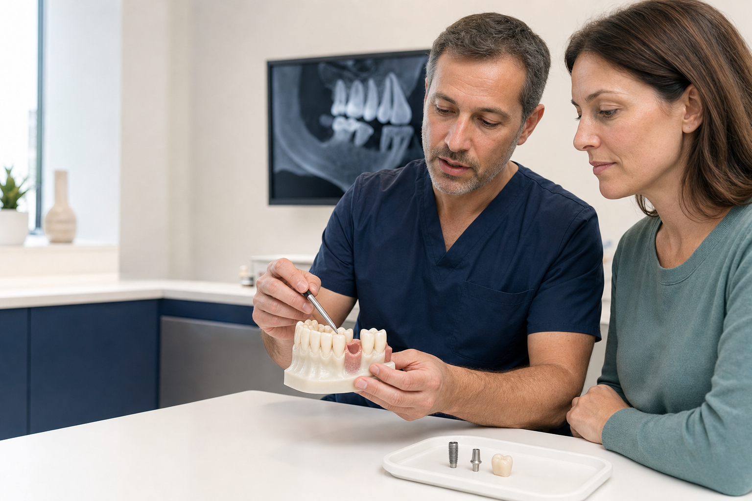
Definition: A single dental implant is a surgically placed fixture supporting one replacement crown. The implant, abutment and crown are separate components with different risks and maintenance needs.
What is a single dental implant?
A dental implant is usually a titanium fixture placed in jawbone to support a prosthetic tooth. After integration, an abutment connects the implant to a crown. Some systems use zirconia components in selected circumstances. An implant does not recreate a natural periodontal ligament and is not immune to inflammation or mechanical failure.
When may it be considered?
A single implant can replace a tooth lost through decay, fracture, trauma, congenital absence or failed treatment. It avoids preparing neighbouring teeth for a conventional bridge. Suitability depends on general health, oral hygiene, bone and soft tissue, space, bite, growth completion and the long-term value of adjacent teeth.
Alternatives
Options include leaving the space, orthodontic closure, a resin-bonded bridge, conventional bridge or removable tooth. A resin-bonded bridge may be especially conservative for some front-tooth spaces. Saving a restorable natural tooth can be preferable to extraction and implant placement; prognosis should be compared rather than assuming an implant is an upgrade.
Assessment and records
The clinician reviews medical conditions, medications, smoking, periodontal history and previous healing. The site is examined for space, gum thickness, bone contour and neighbouring roots. Periapical radiographs and, where justified, CBCT help assess anatomy. A scan or impression allows prosthetically driven planning from the desired crown backward to the implant position.
Why three-dimensional position matters
The implant must be positioned to support a cleanable crown with appropriate emergence and screw access while avoiding nerves, sinuses and roots. Too facial a position can cause recession or grey shine-through; too deep can complicate hygiene; an incorrect angle can force a cemented or over-contoured crown. Guided surgery can transfer a digital plan but cannot compensate for a poor plan.
Bone and soft-tissue requirements
After extraction, bone and gum contours change. Some sites require ridge preservation, guided bone regeneration or a soft-tissue graft. Grafting increases treatment time, cost and morbidity and does not guarantee the original anatomy. Short or narrow implants may be alternatives in selected sites, but evidence and component limitations must be considered.
Timing after extraction
Immediate placement
The implant is placed at extraction. This can reduce surgical episodes but does not prevent ridge remodelling and requires suitable anatomy, infection control and primary stability.
Early placement
Placement occurs after soft-tissue healing or partial bone healing. It can balance tissue management and treatment time.
Delayed placement
The socket heals more fully before surgery. This may be appropriate after infection or major grafting, although ridge shrinkage can increase reconstruction needs.
Loading and the temporary tooth
Immediate loading means a provisional crown or prosthesis is connected soon after placement. It is different from immediate placement. The temporary may be kept out of bite to protect stability. Conventional loading allows healing before connection. Primary stability, bone quality, site and patient factors determine the protocol.
The surgical procedure
Local anaesthesia is commonly sufficient. The surgeon prepares the site and places the implant according to the plan, sometimes with a flap and sometimes flapless. A cover screw or healing abutment is fitted. Sutures may be used. Sedation changes comfort and memory but does not replace local anaesthesia or surgical assessment.
Healing and osseointegration
Bone remodels around the implant over weeks and months. Healing time varies by site, grafting and stability. Patients should avoid loading the area outside instructions, maintain modified cleaning and attend reviews. Pain and swelling usually peak early; increasing symptoms, fever, pus, persistent numbness or implant movement need urgent contact.
Making the crown
After integration, a digital or conventional impression records implant position and tissue. The crown may be screw-retained or cement-retained. Screw retention improves retrievability and avoids subgingival cement, but the access position must be acceptable. Cemented crowns can solve some angulation issues but require meticulous cement control.
Aesthetic-zone challenges
A front implant must harmonise tooth colour, texture, gum margin and papillae. The neighbouring bone largely supports papilla height. A temporary crown may shape tissue gradually. A high smile line, thin tissue, facial bone loss or an unfavourable implant position increases aesthetic risk. Pink ceramic may camouflage tissue loss but does not regenerate it.
How successful are single implants?
A systematic review of studies with at least ten years of follow-up reported implant-level survival around 95%, while survival of the original single crown was lower, around 89.5%. Another review reported technical events such as screw loosening, loss of retention and veneering fracture. Survival is not the same as remaining complication-free.
Early and late failure
Early failure occurs when integration does not establish, sometimes without a single identifiable cause. Late loss may relate to peri-implantitis, overload, fracture or systemic/local factors. If an implant fails, removal, healing, grafting and replacement may be possible, but the site may become more complex.
Peri-implant mucositis and peri-implantitis
Mucositis is inflammation in the soft tissue without progressive supporting bone loss and can often improve with early professional and home plaque control. Peri-implantitis includes inflammation and progressive bone loss. Previous periodontitis, smoking, poor plaque control, inaccessible crown contours and lack of maintenance can increase risk.
Medical and behavioural risk factors
Diabetes control, smoking, antiresorptive or antiangiogenic medication, immune status, radiotherapy and healing history require individual evaluation. Bruxism may increase technical complications. No single risk factor automatically decides treatment, but informed consent and maintenance should reflect the combined profile.
Cleaning a single implant
Brush twice daily and clean the implant contact areas using floss, interdental brushes or other devices selected for the crown contour. Bleeding should not be accepted as normal. Professional reviews include probing where appropriate, tissue assessment, plaque control, bite and radiographs based on risk and baseline comparison.
Implant crown complications
The crown can chip, fracture, loosen, wear the opposing tooth or lose cement. The screw or abutment can loosen or fracture. Food trapping may result from tissue change or poor contact. Retrievable designs facilitate repair, but every component has manufacturer-specific tools and parts.
Implant materials and surface claims
Most established implants are made from commercially pure titanium or titanium alloy with a treated surface. Zirconia implants are available for selected indications but have different component designs and a smaller long-term evidence base. Terms such as “Swiss,” “German,” “premium” or “lifetime” do not identify a device. Ask for the manufacturer, model, regulatory status and published evidence for the actual system.
Diameter, length and site anatomy
Implant dimensions are selected from available bone, tooth space, restorative load and component strength. A wider or longer implant is not automatically better if it compromises surrounding bone. Short implants may avoid grafting in selected posterior sites; narrow implants may fit limited spaces but have indication and strength constraints. Dimensions should be explained in the context of the crown.
Contact points and food trapping
Natural teeth can drift while an integrated implant remains relatively stationary, so the contact beside an implant crown may open over time. Food packing can inflame tissue and damage the neighbouring tooth. The clinician evaluates contact, crown contour and tooth movement. Repair may involve adding material, remaking the crown or treating adjacent tooth position.
Baseline records after restoration
Once the crown is fitted, a baseline clinical examination and radiograph help future comparison. Records should include probing findings where appropriate, tissue condition, crown retention, screw torque and occlusion. Bone levels should be interpreted against this baseline rather than a generic millimetre threshold without chronology.
Implant removal and replacement
A failed or malpositioned implant may be removed with specialised instruments or surgery. Removal can damage bone, and the site may require grafting before another implant. Sometimes a bridge or removable option is safer than repeated surgery. The original treatment warranty may not cover graft reconstruction, provisional teeth or care outside the clinic.
Age and timing
Implants are generally delayed until craniofacial growth is complete because they do not erupt with neighbouring teeth. Even in adults, natural teeth can continue small positional changes, especially in the front region. Older age alone does not prohibit treatment, but medical risk, dexterity, maintenance access and expected future care should be considered.
Understanding the quotation
Confirm whether the fee includes extraction, socket preservation, implant fixture, cover screw, healing abutment, custom abutment, temporary tooth, definitive crown, graft membrane, sedation, imaging and reviews. “Implant price” sometimes covers only the fixture. The final invoice and implant passport should match the components placed.
When to request a second opinion
A second opinion is valuable when a restorable tooth is being extracted, major grafting is proposed, the implant would be close to a nerve or sinus, or the aesthetic risk is high. Share diagnostic-quality radiographs and the proposed crown position, not only a cropped CBCT screenshot. The reviewer should compare tooth preservation, bridge options and implant timing.
Consent should be site-specific
Generic implant consent is not enough. The discussion should identify the site's nerve, sinus, recession, graft, aesthetic and loading risks; the likely temporary tooth; and what happens if primary stability or bone volume differs from the scan. The patient should know which decisions may change during surgery and which require renewed agreement.
Treatment in Turkey
Ask for the implant manufacturer, exact implant and abutment reference, dimensions and component passport. Confirm who performs surgery and restoration, which scans justify the plan and whether grafting is included. Leave time for healing rather than assuming “same day” means definitive completion.
Continuity of care abroad
Verify whether compatible components and drivers are available in your home country. Obtain radiographs, torque records, graft materials, crown retention type and warranty exclusions. Identify a local clinician willing to maintain the implant. A warranty cannot substitute for accessible diagnosis and treatment.
Questions to ask
- Can the natural tooth predictably be saved?
- Why is an implant preferable to a resin-bonded or conventional bridge?
- What bone or soft-tissue graft is expected?
- Is placement immediate, early or delayed—and is loading separate?
- Will the crown be screw- or cement-retained?
- What is the personalised peri-implant maintenance schedule?
Frequently asked questions
Does implant surgery hurt?
Local anaesthesia controls pain during surgery. Postoperative soreness and swelling vary with site and grafting. Severe or increasing pain needs review.
Can an implant get a cavity?
The implant cannot decay, but neighbouring teeth can, and the implant tissues can develop mucositis or peri-implantitis.
Is an implant for life?
No device can be guaranteed for life. Many function long term, but biological and technical maintenance and possible replacement must be anticipated.
Sources and clinical review references
- Hjalmarsson L, et al. Ten-year survival of single implants and crowns. Eur J Oral Implantol. 2016.
- Jung RE, et al. Survival and complications of implant-supported single crowns. Clin Oral Implants Res. 2012.
- Howe MS, et al. Long-term ten-year dental implant survival. J Dent. 2019.
- Wang HL, et al. AO/AAP consensus on peri-implant diseases. J Periodontol. 2025.
Content status: editorial draft for review by a licensed dentist before publication. Last evidence check: 22 July 2026.