
Clinical review required: This evidence-informed draft supports informed discussion. It cannot determine whether a lateral sinus augmentation is appropriate for an individual patient.
What is an open sinus lift?
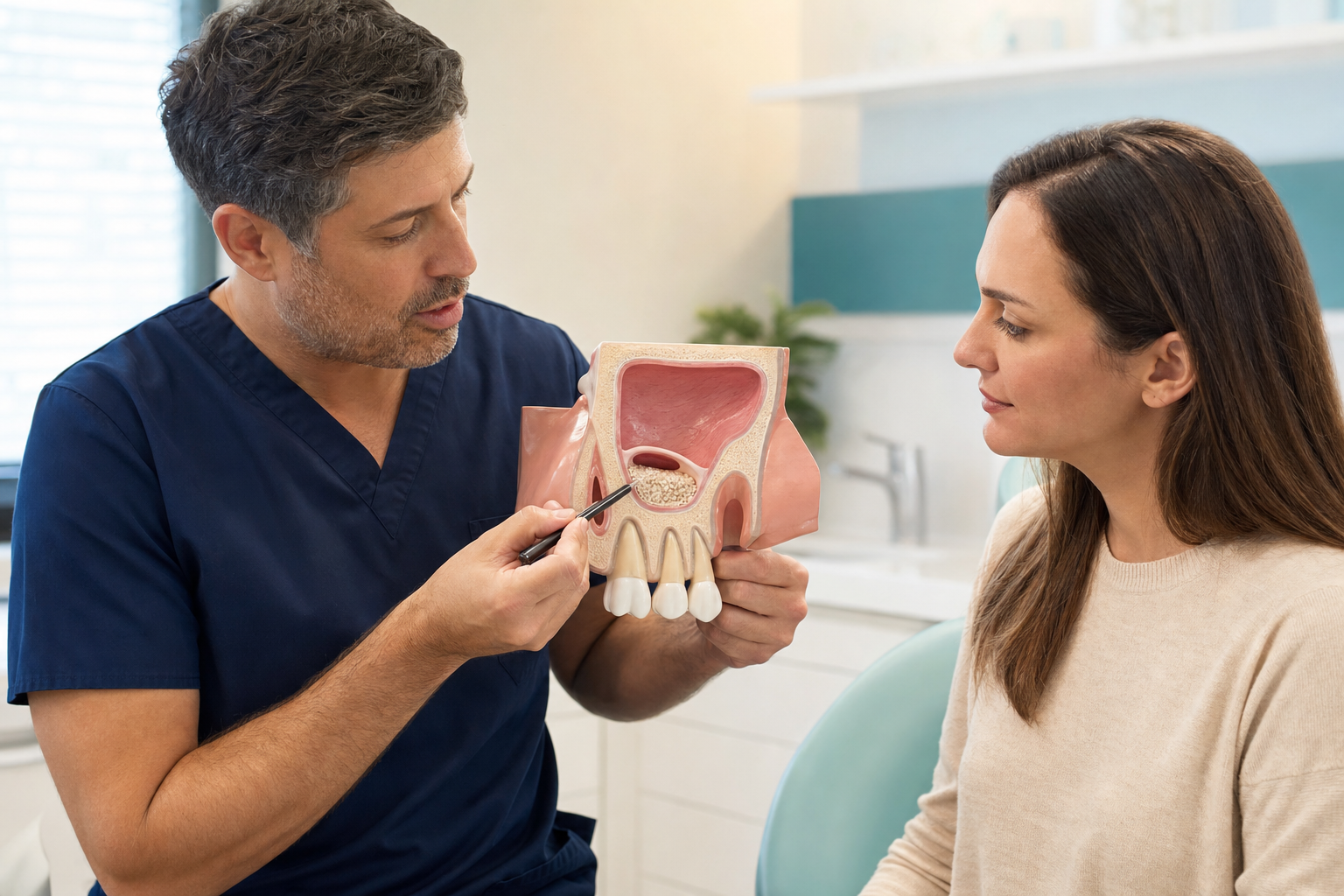
An open sinus lift, usually called lateral-window maxillary sinus floor augmentation, increases bone height in the back of the upper jaw through a window made in the outer sinus wall. The surgeon exposes the Schneiderian membrane, carefully elevates it from the floor and walls, and maintains the new compartment with graft material, a stable clot or both. Implants can be placed simultaneously when native bone provides appropriate stability, or in a second operation after healing.
“Open” describes the lateral access; it does not mean the sinus is left open to the mouth or outside. The wound is closed, and the membrane should separate the augmented space from the sinus cavity. The technique offers greater access and potential volume than a crestal approach but involves a larger operation.
Why is a lateral window used?
The approach is commonly chosen when residual posterior maxillary bone is severely reduced, several adjacent implants require support, a larger vertical gain is planned or sinus anatomy makes a crestal lift unpredictable. Direct access helps the surgeon elevate membrane across a broad area and work around septa. The indication should arise from a prosthetic plan showing where implants need to be, rather than an arbitrary desire to use long implants.
Diagnosis and restorative planning
The proposed crowns determine implant positions, emergence and loading. Clinical assessment covers ridge width, soft tissue, periodontal condition, adjacent teeth, smile and bite. CBCT measures the residual ridge and shows the lateral wall, sinus width, septa, membrane, vascular canals and pathology. A broad sinus can require more graft and may heal differently from a narrow contained space. Planning software assists measurements but does not replace surgical judgement.
Checking sinus health
The maxillary sinus must drain through its natural opening. Active infection, significant obstruction or untreated dental disease can compromise augmentation. Ask about unilateral blockage, facial pressure, discharge, recurrent sinusitis, smell changes, allergies and previous ENT surgery. Mucosal thickening alone is not an automatic contraindication, but symptoms and cause matter. An ENT opinion may be appropriate where imaging or history suggests impaired drainage or unexplained disease.
Sinus septa and vascular anatomy
Bony septa can divide the sinus floor and increase the difficulty of membrane elevation. The window may need modification or more than one access. A vessel within the lateral wall can bleed during window preparation; CBCT may show its canal. Bleeding is usually controllable but can obscure the membrane and increase perforation risk. These details illustrate why a panoramic image alone may be inadequate for complex lateral surgery.
Step-by-step overview
After anaesthesia, the surgeon raises a flap and outlines a window in the lateral maxillary wall with rotary or piezoelectric instruments. The bony plate may be removed, hinged inward or preserved for repositioning. The membrane is detached progressively from the floor and walls without excessive tension. Any perforation is classified and repaired. Graft is placed without overpacking, implants are inserted if planned and stable, a covering membrane may be applied, and the flap is closed without tension.
Rotary instruments versus piezosurgery
Rotary burs are efficient but require controlled handling near the membrane. Piezoelectric devices selectively cut mineralised tissue and are promoted as membrane-protective. A systematic review found the available trials too weak to confirm a meaningful reduction in perforations and suggested slightly longer surgery. Either method can be used safely by trained clinicians. Technology should support visibility and control, not replace sound access and membrane technique.
Graft choices
Options include autogenous bone, allograft, xenograft, alloplast and mixtures. Autogenous bone provides living cells and biological signals but requires a donor site and remodels relatively quickly. Slowly resorbing particles can help maintain space but may remain visible for years. No single material is ideal for every sinus. The volume, healing time, planned implant timing and patient preferences should guide selection. Exact origin, brand and traceability belong in consent and operative records.
Can lateral elevation be graft-free?
Selected lateral procedures use the elevated membrane, repositioned bony window, implants or space-maintaining devices to stabilise a clot without particulate graft. Evidence shows bone formation is possible, but technique and anatomy are critical. Graft-free does not mean risk-free or instantly regenerated. Large unstable compartments may collapse, and the approach should not be chosen solely to market a “biological” package.
One-stage versus two-stage treatment
In one-stage surgery, implants are placed during augmentation. Their initial stability must come from residual native bone. Two-stage treatment grafts first, allows maturation and places implants later. It is useful when native height or quality cannot stabilise implants, infection has been treated or a large reconstruction needs reassessment. Staging increases time and surgery but can reduce the consequences of forcing an unstable implant. The decision should be agreed before the procedure, including contingencies.
Healing time
Augmented bone matures over months. Timing varies with defect size, graft, simultaneous implantation, primary stability and patient biology. Soft-tissue comfort returns long before the graft is ready to support restorative loading. Later imaging and clinical stability guide implant placement or restoration; a standard “three-month teeth” promise is not appropriate for every lateral augmentation.
Membrane perforation
Perforation is the most frequent intraoperative complication. Thin membrane, septa, scars, difficult angles and uncontrolled instruments increase risk. Small tears are commonly covered with a collagen membrane; larger defects may be sutured, patched with layered barriers or lead to postponement. A systematic review reported 97.68% implant survival beneath repaired perforations versus 98.88% beneath intact membranes, without a statistically significant difference, but tear size and sound repair remain clinically important.
What happens if the membrane cannot be repaired?
The surgeon may abandon graft placement, close the site and allow healing before reassessment. Continuing through a large uncontrolled defect can disperse graft into the sinus and increase infection risk. A staged return is inconvenient but may be safer. The consent process should explain this possibility, potential additional costs and who provides follow-up—especially for patients travelling internationally.
Postoperative sinusitis
Sinusitis may present with worsening one-sided pressure, blocked nose, foul discharge, fever or pain after initial improvement. Causes can include infection, obstructed drainage, displaced graft, membrane disruption or an implant problem. Management may involve antibiotics based on clinical judgement, nasal therapy, imaging, drainage, graft or implant removal and ENT collaboration. Repeated remote prescriptions without examining the surgical site can delay definitive care.
Bleeding and bruising
The lateral-wall vessel and soft tissues can bleed. Surgeons use pressure, cautery, bone wax or other haemostatic methods as appropriate. Bruising may extend into the cheek or under the eye. Patients taking anticoagulants or antiplatelet drugs need a coordinated plan but must not stop them independently. Persistent brisk bleeding, dizziness or expanding swelling requires urgent assessment.
Other risks
- Pain, swelling, limited mouth opening and temporary nasal congestion.
- Wound dehiscence, barrier exposure, graft-particle loss or infection.
- Oral-antral communication or chronic sinus symptoms.
- Insufficient bone formation, graft resorption or need for regrafting.
- Implant failure, displacement or an implant position that compromises the crown.
- Damage to adjacent teeth, altered sensation or soft-tissue scarring.
- Donor-site problems when autogenous bone is harvested.
Implant survival and what it means
Meta-analysis has found high survival after both lateral and crestal elevation, with no clear overall difference between techniques. However, survival records whether an implant remains, not whether every sinus stayed symptom-free, the graft was fully vital, the crown is easy to clean or the patient avoided additional surgery. Study heterogeneity and variable follow-up limit individual prediction. Long-term maintenance remains essential.
Recovery experience
Swelling often peaks during the first few days and then declines. Bruising, tightness and temporary nasal stuffiness are possible. Sutures are reviewed or removed according to material. Most daily activity returns before bone maturation, but strenuous work and sport need individual advice. Bilateral surgery and large grafts can create more disruption than a single local window.
Aftercare and sinus precautions
Take prescribed medicines correctly, maintain hydration and use a soft diet. Clean the mouth as instructed without brushing directly over an early wound. Avoid smoking, vaping and manipulation. Do not blow the nose forcefully, and sneeze with the mouth open. Follow restrictions on exercise, swimming, straws, wind instruments and flying. Do not wear an unadjusted denture over the graft.
When urgent assessment is needed
Seek advice for increasing pain or swelling after early improvement, fever, pus, foul nasal discharge, persistent nosebleed, fluid moving between mouth and nose, wound opening, visible large quantities of graft or implant mobility. Rapid facial swelling, eye symptoms, severe headache, breathing difficulty or systemic deterioration warrants urgent or emergency assessment.
Risk reduction
Control dental and periodontal infection, improve plaque control and address nicotine use before surgery. Diabetes and relevant systemic conditions should be stabilised. A complete medication history is necessary, including antiresorptive and antiangiogenic drugs. Careful CBCT planning, tension-free closure and accessible follow-up reduce avoidable problems. Prophylactic antibiotics are prescribed according to case risk and local stewardship; they do not compensate for obstructed sinus drainage or poor surgery.
Alternatives
Short implants, tilted implants, a different distribution of implants, a bridge, removable prosthesis or no replacement may avoid a lateral window. Some alternatives reduce surgery but create different biomechanical or hygiene demands. Short implants are not simply “inferior,” and long implants are not automatically better. Compare the complete restorative pathway, expected maintenance and consequences of failure.
Treatment abroad
Obtain the CBCT assessment, indication, target gain, graft and membrane identities, implant system and staging plan. Confirm access to ENT and surgical care if sinusitis or perforation occurs. Stay for early wound review and suture management. Carry operative notes and traceability information home. Flights and compressed itineraries should follow the surgeon’s case-specific sinus precautions, not a tourism schedule.
Questions to ask
- Why is a lateral window necessary for my restorative plan?
- Is the sinus drainage pathway healthy?
- Are there septa or lateral-wall vessels?
- Which graft and barrier will be used?
- Will implants be placed now or later, and why?
- How are different sizes of membrane tear managed?
- What symptoms trigger ENT involvement?
- Who provides follow-up if I travel home?
Frequently asked questions
Is an open sinus lift major surgery?
It is more extensive than a crestal lift, though it is usually performed as outpatient oral surgery with local anaesthesia and optional sedation.
Does a perforated membrane mean the graft must fail?
No. Many repaired perforations heal successfully, but size, contamination and the quality of repair affect management.
Can both sides be treated together?
Sometimes, but bilateral swelling, procedure time, medical risk and support at home should be considered.
Can I fly immediately afterward?
Usually a buffer is advised because pressure, bleeding and access to care matter. Follow the operating surgeon’s case-specific instructions.
Sources and clinical review references
- Implant survival after direct and indirect sinus elevation.
- Repair of membrane perforations during lateral sinus augmentation.
- Piezoelectric versus rotary instruments for lateral elevation.
- Clinical outcomes after sinus-floor and membrane perforation.
Editorial review note: Evidence reviewed 22 July 2026. Named clinician review is required before indexation.