
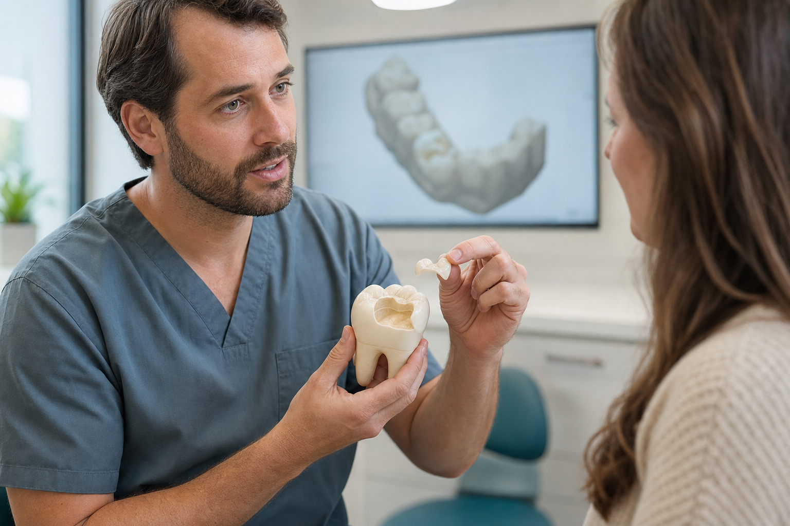
Clinical scope: A dental onlay is a partial-coverage restoration that replaces internal tooth structure and intentionally covers at least one weakened cusp. It can preserve more tissue than a full crown, but only when cracks, margins, pulp, isolation and remaining walls make adhesive partial coverage predictable.
What is a dental onlay?
An onlay restores a posterior tooth and covers one or more cusps judged vulnerable to fracture. It may be placed directly with resin composite or fabricated indirectly from ceramic, composite or metal. The restoration sits within and over selected parts of the tooth rather than wrapping around every surface as a crown generally does.
Why cover a cusp?
Decay, a large old filling, a crack or root-canal access can leave a cusp thin and unsupported. Covering it redistributes contact away from a fragile edge and provides restorative material of controlled thickness. Cusp coverage is not automatically required because a filling is large; thickness, crack direction, bite and remaining marginal ridges inform the decision.
Onlay versus inlay
An inlay stays between the cusps. An onlay extends over at least one cusp. If all cusps are strong, inlay or direct filling may preserve more tissue. If a cusp is compromised, an inlay leaves the central mechanical problem untreated. The final design may change after old material and decay reveal the true structure.
Onlay versus overlay
Terminology varies. An overlay generally covers all cusps or most of the occlusal table while preserving more axial tooth than a full crown. An onlay may cover one or several cusps. Because clinics use labels inconsistently, the patient should view the design and ask which surfaces and cusps will actually be reduced.
Onlay versus crown
A crown usually reduces the tooth circumferentially and covers all cusps. An onlay selectively removes weak tissue and preserves sound walls and enamel for bonding. A crown may provide a more suitable form when damage, cracks, existing crown preparation or retention needs are extensive. “More conservative” is meaningful only when the remaining tooth is genuinely reliable.
Potential indications
An onlay may restore a large posterior cavity, replace a broad defective filling, protect cracked or undermined cusps, rebuild wear or provide cuspal protection after root-canal treatment. The tooth must be restorable, the pulp or endodontic condition controlled and margins accessible. Occlusal space must accommodate adequate material without excessive reduction.
When an onlay may not work
A vertical root fracture, deep circumferential decay, inadequate ferrule-like support, uncontrollable moisture or severe subgingival damage can make partial coverage unreliable. Very short or heavily damaged teeth may need another design. Active periodontal disease and uncontrolled caries are treated first. Bonding cannot make a hopeless tooth predictable.
Cracked tooth assessment
Cracks are evaluated with symptoms, bite tests, magnification, transillumination and removal of old restorations. Their depth and direction may remain uncertain. An onlay can protect selected cusps but cannot heal a crack or guarantee it will not progress. Root involvement, isolated deep probing or persistent pulpal symptoms may worsen prognosis.
Root-treated teeth
Posterior root-treated teeth often have extensive tissue loss and may benefit from cuspal coverage. The access cavity alone is not the only issue; marginal ridges, cracks and existing restorations matter. An onlay or endocrown may preserve tissue in selected cases, while other teeth require a crown and core. A post does not reinforce cusps.
Direct composite onlay
Composite can be placed directly to cover selected cusps in one visit. It is repairable and avoids a laboratory step. Large-volume placement requires controlled adaptation, curing and anatomy. Current evidence supports direct cusp-covering composite in selected teeth but includes fewer long-term studies than some indirect options, so claims should remain measured.
Indirect composite onlay
Fabrication outside the mouth allows polymerisation and contour to be controlled before bonding. Indirect composite may be less brittle and easier to repair than ceramic, but can wear or discolour. Reviews comparing direct and indirect resin techniques have not produced a universal winner, and remaining tooth structure remains central.
Ceramic onlay
Glass-ceramic onlays can provide colour stability, wear resistance and strong adhesive integration when thickness and isolation are suitable. Ceramic is brittle and vulnerable to defects, thin edges and unfavourable contact. Systematic reviews report acceptable medium-term survival, with fracture, debonding, caries and marginal deterioration among recognised outcomes.
Zirconia onlay
Zirconia offers high strength but has different bonding and optical behaviour from glass ceramic. Evidence for partial-coverage zirconia is less mature in some indications. It cannot be etched with hydrofluoric acid like lithium disilicate. Preparation retention, controlled air abrasion and MDP-compatible chemistry may be important according to the system.
Gold onlay
Cast gold can be thin, precisely finished and kind to opposing enamel when polished. It has long clinical history but is visible and requires an alloy-specific laboratory workflow. Cost and patient preference limit use. Metal-free alternatives may be more aesthetic but introduce different thickness, fracture and adhesive considerations.
Choosing which cusps to cover
The dentist assesses remaining width, undermining, cracks and occlusal contact. Functional and non-functional cusp labels alone do not settle the decision. A cusp carrying a heavy contact or separated by a crack may need reduction; a thick sound wall may remain. Reduction guides can prevent arbitrary removal and verify restorative thickness.
Preparation design
Weak tissue and unsupported enamel are removed, internal angles rounded and selected cusps reduced. Ceramic needs adequate bulk and smooth transitions. Adhesive preparations can avoid traditional retentive boxes but still require a stable insertion path and accessible margins. Extremely thin “no-prep” onlay claims may create overcontour or fragile material.
Margin location
Supragingival enamel margins are easier to isolate, scan, bond and maintain. Deep proximal decay may place a margin near the gum. Options include proximal margin elevation, periodontal or orthodontic exposure, a different restoration or extraction. Tissue health and biological width must not be sacrificed simply to keep an adhesive onlay label.
Digital scan and design
An intraoral scan records preparation, adjacent teeth, opposing arch and bite. CAD software helps assess thickness and contact. It cannot detect every crack or automatically choose safe cusp coverage. Scan stitching, moisture and hidden margins can introduce errors. The dentist reviews the proposal rather than accepting default anatomy.
Same-day onlay
Chairside CAD/CAM can mill and place an onlay in one appointment. This avoids a temporary and second anaesthetic, but preparation, scanning, milling, crystallisation or finishing, try-in, bonding and bite adjustment still need adequate time. Same-day describes scheduling, not a different standard of care.
Laboratory-made onlay
A technician fabricates the restoration from an impression or scan and can characterise anatomy and shade. A provisional protects the tooth meanwhile. Laboratory origin does not guarantee fit; the clinician verifies margins, contacts and occlusion. The exact material, block or ingot and surface treatment should be recorded.
Immediate dentine sealing
Freshly prepared dentine may be sealed before impression or scanning in some adhesive protocols. This can support bond development and reduce sensitivity, but requires compatible materials and careful provisional management. It is one part of a controlled workflow rather than a branded guarantee of painless or permanent treatment.
Provisional phase
A temporary onlay seals the tooth and maintains position. It may not reproduce final strength and can loosen. Hard or sticky foods are avoided. Persistent pain during the temporary phase needs diagnosis before definitive bonding. Copying an uncomfortable temporary into ceramic does not solve the underlying problem.
Try-in and verification
The onlay must seat fully without rocking. Margins, contacts, shade, material thickness and internal fit are evaluated. A tight contact can hold the restoration high. Adjusting a thin ceramic edge or connector may create damage. Visible work is approved before bonding while correction remains feasible.
Adhesive bonding
Glass ceramic is etched and silanised; zirconia and composite require their own protocols. Enamel and dentine are conditioned with compatible adhesive under reliable isolation. Resin cement is seated under controlled pressure and excess removed. Mixing surface treatments or contaminating the field can cause early debonding.
Occlusal adjustment
Contacts are checked in maximum closure and excursions after bonding. Heavy force at a margin or over a thin area can initiate failure. Grinding removes glaze or polish, so material-specific repolishing follows. A night guard may reduce parafunctional risk, but it is not a substitute for correct thickness and contact design.
Fracture
Fracture is a common reported failure mode for tooth-coloured onlays. It may involve restoration, cusp or root. Causes include cracks, inadequate thickness, processing defects and overload. A small composite chip can sometimes be repaired; ceramic bulk fracture or a deep tooth crack may require replacement, crown, root treatment or extraction.
Debonding
An onlay can lose retention through contamination, surface-treatment error, preparation limitations or load. The tooth and restoration are inspected before rebonding. If caries or fracture is present, simple recementation is unsafe. Recurrent debonding requires redesign rather than escalation to the strongest available cement.
Marginal deterioration and caries
Margins may stain, wear or gap. Staining alone does not prove active decay. Caries risk, texture, progression and radiographs guide intervention. Fluoride, sugar-frequency control and cleaning remain necessary because the tooth can decay beside ceramic, composite or gold. A repair may preserve tissue when the defect is local.
Pulp complications
Deep decay, cracks and preparation can irritate the pulp. Mild improving sensitivity may be monitored; spontaneous, lingering or worsening pain needs assessment. Root-canal treatment may sometimes be performed through an onlay, but this weakens and alters it. Pulp prognosis should be discussed before expensive definitive fabrication.
Longevity and interpretation
Reviews report acceptable medium-term survival across many direct and indirect tooth-coloured onlays, with broad ranges reflecting different studies. Survival can include repair, rebonding or endodontic treatment. Population figures are not guarantees. Material, tooth vitality, preparation, caries risk, bite and maintenance shape the individual forecast.
Advantages
An onlay can protect weak cusps while preserving sound axial tooth, offer controlled anatomy and remain more repairable or retrievable than some full crowns. Adhesive ceramics can provide excellent aesthetics. These benefits depend on enamel, isolation and design. Partial coverage is not a goal when it leaves a dangerous crack or inaccessible margin.
Limitations
The technique is sensitive, can require two visits and may cost more than a direct filling. Ceramic can fracture; resin can wear; any material can debond or develop marginal caries. Deep margins complicate bonding. Future replacement removes additional tissue, so the initial indication should be strong and documented.
Aftercare
Brush twice daily with fluoride toothpaste and clean proximal margins. Avoid chewing hard objects and report a changed bite, movement, fracture, floss shredding or persistent symptoms. Reviews inspect remaining cusps and crack progression as well as the restoration. A serviceable onlay is not replaced solely because of age.
Treatment abroad
Request preoperative records, exact material, cusp-coverage design, scan or impression, bonding system and laboratory traceability. Allow time for provisional assessment, remake and bite review. Ask who will manage a pulp flare-up or debond after travel. A manufacturing warranty is not the same as biological follow-up.
Questions to ask
- Which cusps need coverage and why?
- Is there a crack and how deep may it be?
- Why not a direct filling or full crown?
- Which exact material is planned?
- Can the margins be isolated?
- What happens if symptoms persist?
Frequently asked questions
Is an onlay better than a crown?
It preserves more tissue in suitable teeth, but a crown may be more appropriate when damage is extensive. Neither label is universally better.
Does an onlay protect a cracked tooth?
It can protect selected cusps, but cannot guarantee a crack will stop or compensate for root involvement.
Can an onlay be repaired?
Some resin and local ceramic defects can be repaired. The material, damage and underlying tooth determine feasibility.
Sources and clinical review references
- Survival of direct composite and indirect tooth-coloured adhesive onlays.
- Longevity of ceramic onlays: systematic review.
- Ceramic and resin partial-coverage restorations: meta-analysis.
- Complications and survival of inlays, onlays and crowns.
Editorial review note: Evidence reviewed 22 July 2026. Named clinician review is required before indexation.