
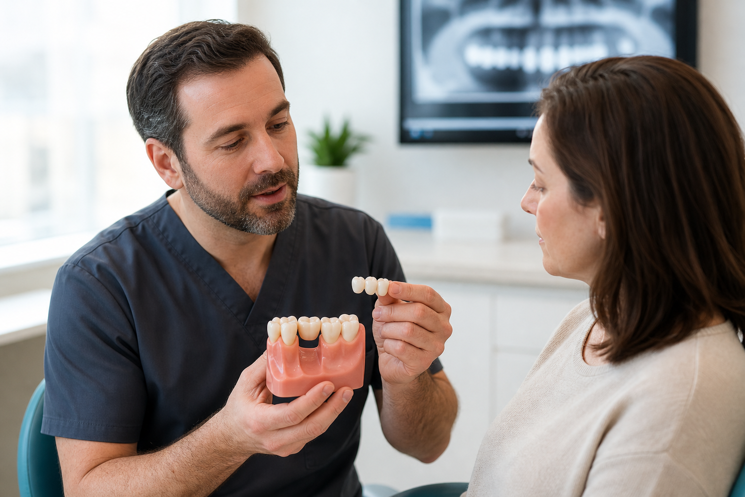
Clinical scope: A dental bridge is a fixed prosthesis that replaces one or more missing teeth. This guide focuses on tooth-supported bridges. Implant-supported bridges have different biological risks and are covered separately. Suitability depends on the missing-tooth space, abutment teeth, bite, hygiene and alternatives—not on the gap alone.
What is a dental bridge?
A conventional dental bridge joins an artificial tooth, called a pontic, to crowns or retainers on neighbouring teeth. The patient does not remove it. A bridge can restore appearance, chewing contact and tooth position, but it transfers force through its supporting teeth. It therefore treats a space by involving teeth on either side or, in selected cantilever designs, one side.
Parts of a bridge
The pontic occupies the missing-tooth space. Retainers attach the bridge to prepared teeth, resin-bonded wings or implants. Connectors join these components. Abutments are the supporting teeth or implants. Each part affects strength and cleaning. A natural-looking pontic that presses too tightly into the gum may be biologically inferior to a slightly more cleansable contour.
Why replace a missing tooth?
Replacement may improve chewing, speech, appearance and stability of adjacent teeth. It is not automatically required for every space. Some shortened dental arches function satisfactorily, while visible or strategically important gaps cause meaningful problems. The decision considers patient priorities, opposing teeth, future movement, periodontal health and whether treatment would sacrifice otherwise healthy tissue.
Conventional fixed bridge
A conventional bridge usually requires substantial preparation of abutment teeth for crowns. It can be logical when those teeth already have large restorations or need crowns independently. Preparing intact teeth solely to replace one missing tooth creates an irreversible cost. The clinician weighs that cost against an implant, resin-bonded bridge, removable appliance or monitored space.
Cantilever bridge
A cantilever bridge has support on one side of the pontic. Conventional cantilevers can create leverage and require cautious use. By contrast, a single-retainer resin-bonded cantilever is an established anterior design because it lets the pontic and one abutment move together. The word cantilever therefore does not describe one uniform risk; support type, span and loading matter.
Resin-bonded bridge
A resin-bonded or adhesive bridge uses a wing bonded mainly to enamel, often with minimal preparation. It preserves more tooth tissue than full-coverage retainers. It is particularly useful for selected single anterior spaces. Bonding area, enamel quality, occlusion and moisture control are decisive. Maryland bridges are discussed in a dedicated guide.
Implant-supported bridge
An implant-supported bridge connects pontics to dental implants rather than natural abutment teeth. It may replace several teeth without preparing neighbours, but introduces surgery, peri-implant disease risk and implant-specific maintenance. Natural teeth and implants differ in mobility, so joining them requires special justification. This page does not substitute for implant assessment.
Who may be suitable?
A candidate needs a restorable space, stable oral disease and abutments capable of carrying additional load. Gum condition, root length, remaining tooth structure, endodontic status, mobility and alignment are evaluated. Expectations must fit anatomy. A bridge cannot restore lost gum volume perfectly or correct severe ridge collapse without additional treatment or a deliberate prosthetic compromise.
Abutment tooth assessment
Each proposed support is examined clinically and radiographically. Decay, cracks, previous root treatment, post design, crown-to-root relationship and periodontal attachment influence prognosis. A large healthy-looking crown above the gum does not guarantee a strong root. If one abutment fails, the connected prosthesis may be lost, making weak-link analysis essential.
Span length and biomechanics
Longer spans flex more and place greater demand on connectors and abutments. Multiple missing teeth, heavy bites and curved arch positions amplify complexity. Adding supports does not automatically solve the problem, because misaligned or mobile teeth may distribute force poorly. The bridge design should be based on load, material dimensions and periodontal support rather than a simple tooth count.
Occlusion and parafunction
The dentist records contact in closure and jaw movements. A pontic or connector exposed to unfavourable lateral force may chip or loosen retainers. Bruxism is not always a contraindication, but it changes material, connector, contact and protective-appliance decisions. A night guard can reduce some mechanical risk; it cannot compensate for an under-designed bridge or untreated mobility.
Gum and bone contour
After extraction, the ridge commonly changes shape. A standard tooth form may appear too long, leave a black space or trap food. Pontic design can create the illusion of emergence in selected anterior sites, while posterior designs prioritise access. Ridge augmentation may improve contour but adds surgery and healing. Expectations should be tested before definitive fabrication.
Pontic shapes
Modified ridge-lap pontics balance appearance and cleansability. Ovate pontics extend into a shaped soft-tissue site for a stronger emergence effect but require healthy, maintainable tissue. Hygienic pontics sit away from the ridge in selected non-visible areas. A saddle-like pontic wrapping broadly over the gum tends to be difficult to clean and is generally undesirable.
Material options
Metal-ceramic bridges combine a metal framework with veneering porcelain. Zirconia frameworks or monolithic zirconia avoid a conventional metal coping. Other ceramics have narrower span and thickness indications. Material choice depends on location, span, connector dimensions, substrate colour, laboratory system and repair strategy. “Metal-free” alone is not a mechanical specification.
Metal-ceramic bridges
Metal-ceramic fixed dental prostheses have extensive clinical history and can provide strong frameworks with controlled dimensions. Veneering porcelain may chip, metal can influence opacity and a margin may become visible with recession. These are design considerations rather than proof the category is obsolete. Alloy selection, framework support and laboratory execution affect outcome.
Zirconia and monolithic ceramic
Zirconia can be layered, micro-layered or largely monolithic. Reducing unsupported veneering ceramic may reduce one chipping pathway, but connectors and occlusal thickness remain critical. Different zirconia generations trade translucency and strength. Recent systematic reviews report favourable performance for several monolithic restorations while noting limited direct comparative evidence for some multi-unit indications.
Preparation and tooth preservation
Conventional retainers require enough reduction for material thickness, a clear finish line and resistance form. Over-preparation risks pulp injury and weakens the tooth; under-preparation can cause bulky contours or thin ceramic. Reduction guides and magnification help. Existing restorations and decay may alter the final preparation beyond what a marketing animation shows.
Impressions and digital scans
Conventional impressions and intraoral scans can both produce accurate work when margins and tissue are controlled. Digital scanning does not make hidden blood or saliva disappear. The laboratory needs the preparation, opposing arch, bite, shade, pontic tissue and provisional information. Long spans may require additional verification because small capture or stitching errors can accumulate.
Provisional bridge
A temporary bridge protects prepared teeth and allows assessment of comfort, bite, speech, contour and cleaning. It can condition anterior tissue. It is not merely cosmetic packaging while the laboratory works. Persistent pressure, looseness or sensitivity during the provisional phase should be investigated rather than copied into the final prosthesis.
Try-in and fit
The clinician evaluates complete seating, marginal integrity, contacts, connector space, shade and pontic pressure. A bridge must seat passively on all abutments; forcing one retainer down can create stress elsewhere. Radiographs may assist selected margin checks. Patient approval of visible form occurs before definitive cementation when correction remains possible.
Cementation
Cement depends on material, preparation retention and moisture conditions. Some bridges use conventional cement; others require adhesive protocols. Excess cement is removed, especially below the gum. Stronger cement cannot rescue major misfit or inadequate preparation. If a bridge will not seat predictably, identifying the obstruction is safer than increasing pressure.
What can go wrong?
Biological complications include decay, pulp inflammation, root fracture and periodontal breakdown. Technical complications include loss of retention, ceramic chipping, connector fracture and wear. Food trapping or speech problems can arise from contour. “Survival” in studies may include a bridge that needed repair, so patients should distinguish survival, success and maintenance burden.
Decay under a bridge
The prosthetic material cannot decay, but abutment tooth margins can. Caries risk rises with plaque, frequent sugars, dry mouth and inaccessible contours. Early disease may be difficult to see beneath retainers. Fluoride, radiographic review when indicated and meticulous margin cleaning matter. Recurrent decay can require removal of the entire bridge even when only one support is affected.
Pulp and root-canal risk
Preparation of a vital tooth may cause temporary sensitivity or later pulp disease. Deep pre-existing restorations increase uncertainty. Root-canal treatment is not routinely required before a bridge, and preventive treatment removes healthy tissue. If treatment becomes necessary later, access may sometimes be made through the retainer; prognosis depends on the tooth and bridge condition.
Chipping, fracture and debonding
Small porcelain chips may be polished or repaired with composite. Larger framework or connector fractures commonly require replacement. A loose retainer allows leakage and should be assessed promptly. Recementation is appropriate only after examining decay, fit and the reason for loss. Repeated debonding signals a design, preparation or load problem.
Cleaning beneath the pontic
A normal toothbrush cannot pass through connected units. Super floss, floss threaders, interdental brushes or an oral irrigator can clean under the pontic and beside abutments. The correct tool depends on space and tissue. An irrigator can help but does not always replace mechanical plaque disruption. The dental team should demonstrate access before cementation.
How long does a bridge last?
No single lifespan applies. Studies estimate group survival over defined periods, not an expiry date for an individual bridge. Abutment health, span, material, caries risk, bite, smoking, hygiene and maintenance influence outcome. A bridge can remain functional for many years, need repair earlier or fail through an unrelated change in a supporting tooth.
Bridge versus implant
A bridge avoids implant surgery and may be efficient when neighbouring teeth need crowns. An implant avoids preparing intact neighbours but requires adequate bone, healing and lifelong peri-implant care. Treatment duration, cost, anatomy and complication type differ. Neither option is universally more conservative: conservation must include teeth, bone, surgery and future retreatment.
Bridge versus removable partial denture
A removable denture can replace several dispersed teeth and lost tissue with less tooth reduction, usually at lower initial cost. It is bulkier and must be removed for cleaning. A fixed bridge feels more tooth-like but may require more support and is less adaptable if other teeth are lost. Future dental trajectory matters.
Bridge versus leaving the space
Monitoring can be reasonable where function and appearance are acceptable and movement risk is low. It avoids immediate intervention but needs review. Teeth do not invariably drift in the same way or at the same rate. The decision should be recorded with expected benefits and consequences, rather than presenting replacement as compulsory.
Treatment abroad
Ask for diagnostic records, abutment prognosis, exact material, pontic and connector design, laboratory traceability and a provisional phase. Allow enough time for tissue review and remakes. Obtain radiographs and cement details for future care. A guarantee may cover fabrication but not biological failure of an abutment, travel or local corrective treatment.
Questions to ask
- Why is a bridge preferable to an implant, adhesive bridge or no replacement?
- Are the supporting teeth healthy enough?
- Which material and pontic form are planned?
- How much tooth must be removed?
- How will I clean beneath it?
- What happens if one abutment fails?
Frequently asked questions
Can a bridge replace two missing teeth?
Sometimes, but span, position, abutment support and material dimensions must be assessed. A longer bridge is not simply a wider version of a short one.
Does food get under a bridge?
The space must allow cleaning. Persistent trapping may indicate unsuitable contour, open contact, tissue change or technique and deserves review.
Do bridge teeth need root canals?
Not routinely. Vital teeth are preserved when healthy, although preparation carries a later pulp risk.
Sources and clinical review references
- Metal-ceramic, veneered and monolithic all-ceramic multi-unit fixed dental prostheses: systematic review and meta-analysis.
- Survival and complications of monolithic ceramic tooth-supported restorations.
- Zirconia-ceramic versus metal-ceramic posterior multi-unit fixed dental prostheses.
- Clinical procedures and designs for all-ceramic resin-bonded fixed dental prostheses.
Editorial review note: Evidence reviewed 22 July 2026. Named clinician review is required before indexation.