
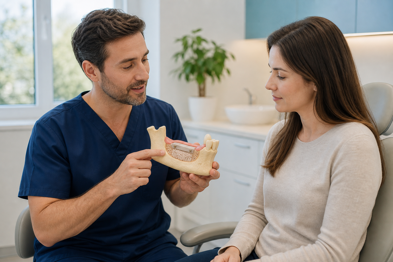
Clinical scope: Dental bone regeneration is a biological and surgical process that creates protected space for new vital bone to form. Guided bone regeneration usually combines defect preparation, graft or blood clot, a barrier membrane, fixation and stable soft-tissue closure. It is not synonymous with adding particles alone.
What is bone regeneration?
Regeneration aims to restore living bone architecture and function rather than merely occupy a space. Cells and blood vessels enter a stable protected defect and deposit new matrix that remodels over time. Biomaterials can guide or support this process. Clinical success is usable bone in the right position, not simply radiopaque graft on a scan.
What is guided bone regeneration?
Guided bone regeneration, or GBR, places a membrane between the bony defect and faster-growing soft tissue. The barrier protects a space where bone-forming cells can populate. Graft particles may support volume, and pins, screws or mesh may stabilise the construct. Membrane choice follows defect shape and required rigidity.
Bone grafting versus bone regeneration
Bone grafting describes placement of material. Regeneration describes the biological outcome and often a wider protocol. A socket filled with slow-resorbing particles may preserve contour without fully replacing every particle with living bone. A well-stabilised clot beneath a membrane can regenerate selected contained defects with little graft.
Why bone is lost
Extraction causes predictable ridge remodelling. Periodontitis, trauma, infection, implant failure, cysts and long-term tooth loss can create larger defects. Thin facial bone is particularly vulnerable. The pattern—contained, horizontal, vertical or combined—matters more than a single width measurement because it determines blood supply and space stability.
Regeneration around natural teeth
Periodontal regenerative procedures can treat selected intrabony defects using membranes, grafts, enamel-matrix products or biologics. Their anatomy and evidence differ from implant ridge augmentation. This guide focuses mainly on edentulous ridges and peri-implant defects; a generic “bone regeneration” label should state the actual target.
Regeneration before implants
A staged GBR procedure can rebuild a ridge before implant placement. This allows healing and confirmation of volume but adds surgery and time. It may be chosen when the defect is too large to stabilise an implant in the correct position or when simultaneous placement would expose threads beyond a predictable regenerative envelope.
Simultaneous GBR
An implant and graft are placed together when the implant can achieve primary stability and the defect is manageable. The implant helps define the needed envelope, but the graft must remain stable and covered. Simultaneous care reduces stages; failure can affect both augmentation and implant.
Contained versus non-contained defects
A defect with remaining bony walls has blood supply and natural space support. A broad horizontal or vertical defect lacks walls and needs more rigid containment. The same membrane and particulate graft should not be expected to perform identically in both. Defect classification guides material, fixation and prognosis.
The PASS principles
Predictable regeneration is often described through primary closure, angiogenesis, space maintenance and stability. These principles are interdependent. A biologically active graft that moves or exposes can fail; a rigid barrier without blood supply cannot create healthy bone. The acronym is a planning framework, not a guarantee.
Blood supply and angiogenesis
New bone depends on vessels from recipient bone and soft tissue. Decortication or small perforations may encourage bleeding in selected protocols, although technique varies. Large blocks and vertical spaces face longer diffusion distances. Smoking, radiation and vascular disease can impair supply and require risk modification or alternative plans.
Space maintenance
Soft tissue exerts pressure and can collapse a flexible membrane into the defect. Graft, tenting screws, titanium reinforcement or mesh preserves a three-dimensional space. Too much rigidity can complicate closure or removal. The desired space must correspond to the prosthetic implant position rather than arbitrary ridge bulk.
Wound stability
Micromovement disrupts vascular ingrowth and clot organisation. Fixation pins, screws or a stable contained socket can immobilise the graft and membrane. An unstable denture or tongue pressure may disturb healing. Patients should understand why they must avoid manipulating the wound even when it feels bulky.
Primary closure
Tension-free closure protects most GBR sites. Flap release advances tissue but can reduce vestibular depth or alter keratinised mucosa. Overfilling makes closure harder. Some socket protocols intentionally heal with a plug or membrane exposed, but that is different from unintended exposure of a large augmentation.
Resorbable membranes
Collagen and other resorbable barriers break down and usually avoid a second removal surgery. They handle well but offer limited rigidity and can lose function quickly if exposed or infected. Cross-linking can alter degradation and tissue response. Fixation may still be required despite the membrane being resorbable.
Non-resorbable membranes
Dense PTFE, expanded PTFE and titanium-reinforced barriers provide longer space maintenance. Many require planned removal. Exposure management varies by surface and contamination. They can support larger defects but demand careful soft-tissue closure and patient compliance. “Non-resorbable” is a material category, not one complication rate.
Titanium mesh
Mesh can maintain complex ridge contours and vertical space. It is shaped and fixed, often with particulate graft. Exposure is possible, and removal is typically required. Sharp edges, soft-tissue thickness and screw position matter. Mesh is not an invisible scaffold that can be left without a retrieval plan.
Graft materials
Autograft supplies cells and signals; allograft, xenograft and alloplast mainly provide scaffolds with different remodelling. Mixtures can balance biological turnover and volume stability. Product selection should match defect and membrane. No particle can compensate for motion, contamination or inadequate blood supply.
Biologics
Growth factors and platelet concentrates may support healing in selected protocols. Their carrier, dose, indication and adverse effects matter. They do not replace space maintenance in a non-contained defect. Claims of “regrowing bone naturally” should identify the product, regulatory status and human clinical evidence.
Recipient-site preparation
Granulation tissue is removed and bone surfaces assessed. Perforations, cortical decortication or marrow exposure may be performed based on technique. Anatomy near nerves and sinus limits manipulation. A clean bleeding recipient bed supports healing, but excessive removal sacrifices the bone the procedure seeks to rebuild.
Digital planning
CBCT and prosthetic wax-up locate the future implant and measure the desired bone envelope. Digital guides or custom meshes may translate the contour. Scan artefact and manufacturing accuracy remain relevant. Planning should include fixation screw and flap location, not only a colourful volume overlay.
Healing stages
The initial clot organises into vascular tissue, woven bone forms and later remodels into lamellar structure. Graft particles resorb at material-specific rates. Radiographic density can reflect residual mineral rather than mature bone. Healing time is selected by defect and protocol, often several months.
Membrane exposure
Exposure allows oral bacteria to reach the barrier and graft. Systematic review evidence shows significantly worse bone gain when membranes expose. Small exposure may be managed with hygiene and monitoring; infection, mobility or large non-resorbable exposure can require early removal. Immediate reporting improves options.
Infection
Drainage, progressive swelling, bad taste, fever or wound breakdown can indicate infection. Antibiotics may support management but cannot stabilise a mobile contaminated construct. Partial or complete graft removal may be necessary. Retreatment occurs only after cause, soft tissue and patient risk are addressed.
Graft particle leakage
A few particles can appear during early socket healing, but ongoing loss with an opening warrants review. Patients should not pick or rinse aggressively. Particle loss can reduce contour without destroying the whole procedure. Clinical stability and tissue coverage determine whether intervention is needed.
Soft-tissue complications
Flap advancement can alter vestibule, papilla and keratinised tissue. Scar, recession or thin coverage can affect implant aesthetics. Soft-tissue grafting may be staged or combined. Bone volume alone does not create a natural emergence profile; the prosthesis and mucosal envelope need joint planning.
Nerve and sinus risks
Fixation screws, graft and instrumentation must avoid mandibular nerves, mental foramen and sinus. CBCT helps map anatomy. Altered sensation, sinus membrane perforation or sinusitis can occur depending on site. More augmentation is not safer when it encroaches on protected structures.
How success is assessed
Success includes uncomplicated healing, sufficient bone for implant placement and stable contour. CBCT measures volume but does not directly prove vitality. Re-entry allows tactile assessment and occasionally a core sample. Implant stability and long-term marginal bone provide later functional evidence. A single postoperative scan is incomplete proof.
How much bone can be regenerated?
There is no universal millimetre promise. Horizontal contained gains are generally more predictable than major vertical augmentation. Defect anatomy, closure and technique cause wide variation. Published averages should not be converted into guaranteed individual dimensions, especially when studies use different imaging and re-entry methods.
Alternatives
Short or narrow implants, tilted placement, a bridge, removable prosthesis, orthodontic movement or no replacement can sometimes avoid major GBR. Each has different compromises. Regeneration should support a meaningful restorative plan, not become an end in itself because a clinic has a preferred membrane system.
Aftercare
Avoid smoking, manipulation, pressure from dentures and hard food at the site. Use prescribed rinses and medicines and clean adjacent areas as instructed. Report opening, exposed membrane, drainage, fever or increasing pain. Scheduled wound checks are as important as the later CBCT because early soft-tissue failure can change outcome.
Treatment abroad
Request defect classification, target gain, exact graft and membrane, fixation plan, implant timing and exposure protocol. Obtain operative notes and product lot details. Remain for early wound and suture reviews. Clarify where non-resorbable membrane or mesh will be removed and who treats complications after return.
Why primary closure and blood supply matter
A barrier can protect a regenerative compartment only when the surrounding tissues can nourish and cover it. Surgeons release and advance flaps to obtain closure without excessive tension while trying to preserve blood supply and vestibular anatomy. Over-thinning, repeated surgery, scar tissue and a large vertical demand can make closure harder. The blood clot beneath the membrane must remain stable long enough for vessels and bone-forming cells to enter. Micromovement, early contamination or collapse reduces the protected space. This is why a sophisticated membrane cannot compensate for poor flap design, unstable fixation or inadequate soft tissue.
Staged versus simultaneous implant placement
Minor dehiscence-type defects may be regenerated around an implant that already has adequate primary stability in a prosthetically correct position. Larger horizontal or vertical deficiencies often require staged regeneration followed by implant placement after healing. Simultaneous treatment can shorten the pathway but should not force an implant into an unstable or compromised position. Staging adds time and another operation, yet it allows direct inspection of the regenerated ridge and may simplify implant placement. The choice depends on residual bone, defect morphology, soft tissue, infection control and the consequences of graft failure.
Maintenance after the implant is restored
Regeneration is not the endpoint. The final crown or bridge must permit cleaning, healthy tissue contours and manageable force. Professional reviews monitor plaque, probing findings, bleeding, suppuration and radiographic bone levels. Smoking, uncontrolled periodontal inflammation and inaccessible prosthetic contours can threaten both native and regenerated bone. Patients need site-specific brushes or interdental aids and a recall interval based on risk. A technically successful augmentation can still lose value if the restoration cannot be maintained.
Questions to ask
- Is the defect contained, horizontal or vertical?
- What bone envelope does the implant actually need?
- How will space and membrane be stabilised?
- Is primary closure expected?
- What happens if the membrane exposes?
- Could another restorative plan avoid major regeneration?
Frequently asked questions
Is regenerated bone the same as graft material?
No. Regenerated bone is living tissue; graft may remodel, persist as scaffold or combine with new bone.
Must a membrane be removed?
Resorbable membranes usually do not; non-resorbable barriers and meshes commonly require planned removal.
Can exposed membrane still succeed?
Sometimes partially, but exposure significantly worsens average outcomes and requires prompt professional management.
Sources and clinical review references
- Guided bone regeneration for implant-related bone augmentation.
- Effect of membrane exposure on guided bone regeneration.
- Complications of resorbable and non-resorbable GBR membranes.
- Alveolar ridge-preservation interventions and bone remodelling.
Editorial review note: Evidence reviewed 22 July 2026. Named clinician review is required before indexation.