
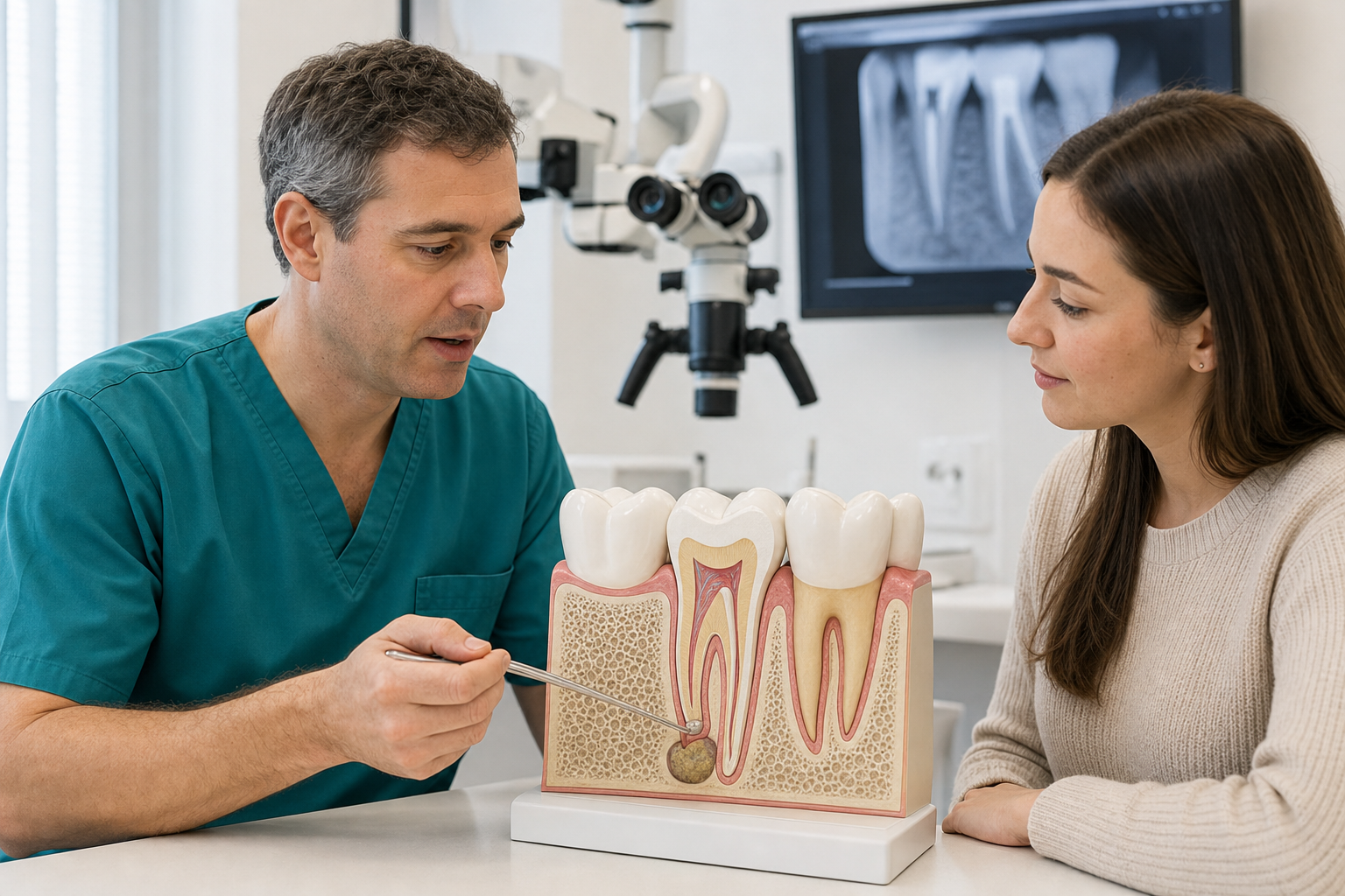
Clinical scope: Apicoectomy is endodontic microsurgery performed through the gum and bone to remove a root tip, treat diseased apical tissue and seal the canal from the root end. It is usually considered for a previously root-treated, restorable tooth when observation or nonsurgical retreatment is unsuitable or has not resolved disease.
What is an apicoectomy?
An apicoectomy, also called root-end resection, exposes the tip of a tooth root through a small surgical access. Inflamed tissue is removed, a short portion of the root is resected, the canal end is prepared and a biocompatible filling is placed. The aim is to preserve the tooth while controlling apical disease.
Apicoectomy and endodontic microsurgery
Traditional apicoectomy may describe root-tip removal alone. Contemporary endodontic microsurgery combines magnification, illumination, small osteotomy, ultrasonic root-end preparation and calcium-silicate or other validated filling materials. These components improve inspection and sealing. Patients should ask which approach is planned rather than relying on the procedure name.
Why might surgery be needed?
Persistent apical periodontitis can result from inaccessible anatomy, apical ramifications, extraradicular infection, leakage, foreign material or a problem not correctable from above. A crown or post may make re-entry destructive. Surgery can directly inspect the root end, remove tissue for biopsy and seal the apical canal.
Is every persistent lesion an indication?
No. Some lesions continue healing for years, and stable scar tissue can remain radiographically. New symptoms, enlargement, a clear apical cause or failed adequate follow-up strengthen the indication. A non-endodontic lesion or root fracture will not be cured by routine apicoectomy, so diagnosis precedes surgery.
Diagnostic assessment
The clinician reviews previous endodontics, symptoms, periodontal probing, crown seal and restorability. Periapical radiographs and selected limited-field CBCT show lesion, cortical bone, roots and nearby structures. CBCT can reveal complexity but has artefacts and radiation. It cannot rule out every vertical fracture.
Apicoectomy versus nonsurgical retreatment
Retreatment re-enters the crown and can disinfect missed canals and leakage along the full system. Surgery treats the root end without necessarily correcting coronal contamination. Comparative evidence is limited and heterogeneous, so neither is universally superior. The likely cause, post removal risk, anatomy and restoration determine the logical route.
Apicoectomy versus extraction
Surgery preserves the natural tooth and avoids an immediate replacement decision. Extraction removes the tooth and lesion source but may require implant, bridge, denture or monitored space. Prognosis, periodontal support, crack risk, cost, recovery and adjacent teeth are compared. A failed surgery can still lead to extraction later.
Who performs the procedure?
Endodontists commonly perform apical microsurgery; oral surgeons and appropriately trained dentists may also operate depending on scope and region. Training with magnification, microsurgical instruments and root-end materials matters. Referral may be particularly important near nerves, sinus, multiple roots or previous surgery.
When surgery may not be suitable
Vertical root fracture, severe periodontal attachment loss, non-restorable decay, unfavourable root length, inaccessible surgical anatomy or significant medical risk may rule it out. A lesion caused by a missed coronal canal may be better treated nonsurgically. Patient ability to maintain and restore the tooth also matters.
Medical and medication review
The team reviews anticoagulants, antiplatelets, diabetes, immune conditions, allergies, smoking and bone-modifying drugs. Medicines should not be stopped without the prescribing clinician’s plan. Blood pressure and infection signs are assessed. Local haemostatic methods manage many patients safely, but individual risk determines setting and coordination.
Local anaesthesia and sedation
Local anaesthetic numbs the surgical area and often includes a vasoconstrictor to control bleeding. Pressure and vibration may still be felt. Conscious or IV sedation can be considered for anxiety or complexity, but adds fasting, escort and monitoring requirements. Sedation does not replace local anaesthesia.
Flap design
A small incision and tissue flap expose the bone over the root. Design depends on tooth, gum margin, blood supply, papilla and existing crowns. The goal is access with minimal tissue trauma and predictable closure. Incisions through poor tissue or close to restoration margins can increase recession or scarring risk.
Bone access
A small osteotomy is made to reach the root end and lesion. CBCT, landmarks and magnification help orientation. Removing excessive bone increases recovery and may affect neighbouring structures; too little access restricts inspection. In selected complex cases, a digital guide may assist, but evidence and accuracy limitations remain.
Removing diseased tissue
Inflamed or cyst-like tissue is curetted from around the root. It may be sent for histopathology, especially when appearance, size or behaviour is atypical. Pathology can confirm inflammatory tissue or identify another lesion. Discarding unexpected tissue without consideration can miss a non-endodontic diagnosis.
Why histopathology can matter
Most tissue removed from an apical lesion is inflammatory, but radiographs cannot identify every lesion type reliably. A specimen can distinguish a radicular cyst, granulomatous inflammation and less common non-endodontic disease. The surgeon should preserve representative tissue and provide the pathologist with tooth, site, symptoms and imaging information. A report that does not match the expected diagnosis may trigger wider investigation or referral. Patients should know whether pathology is included in the fee, when the result will return and who is responsible for discussing it.
Root-end resection
Approximately a few millimetres of the apex may be removed, depending on anatomy and plan, to eliminate apical ramifications and expose the canal cross-section. The cut angle is kept conservative in microsurgery. Excessive resection shortens the root; insufficient access may leave untreated anatomy. The resected surface is inspected for cracks and canals.
Root-end preparation
Ultrasonic microtips prepare a small cavity along the canal axis from the root end. This aims to retain tooth while creating depth for a seal. Preparation must avoid perforation, cracks and thinning. Magnification helps identify isthmuses and multiple canals that a simple round bur preparation could miss.
Root-end filling
Calcium-silicate materials such as MTA-family or bioceramic products are commonly used because they can seal in a moist surgical environment and are tissue compatible. Other materials have evidence. Placement, adaptation and haemostasis matter as much as brand. The filling seals the prepared end; it does not repair coronal leakage.
Microscope and illumination
High magnification and coaxial light improve inspection of the resected root, isthmuses, cracks and filling adaptation. Meta-analytic evidence generally favours contemporary microsurgical protocols over older approaches, although several components change together. A microscope cannot turn a vertically fractured root into a restorable one.
Bone grafting
Many small lesions heal without graft material. Larger through-and-through defects, cortical loss or combined periodontal lesions may prompt use of graft and membrane. Evidence varies by defect. Grafting adds material, cost and potential complications and should address a defined regenerative need rather than be routine upselling.
Sutures
The flap is repositioned and secured with fine sutures. Pressure helps stabilise the clot. Sutures are removed at the interval specified by the clinician, often within days. Plaque control and gentle cleaning support early healing. Pulling the lip repeatedly to inspect the site can disturb closure.
How long does surgery take?
Time depends on tooth, root count, lesion, access, grafting and previous surgery. A single accessible anterior root differs from a molar near sinus or nerve. Scheduling should include consent, anaesthesia, haemostasis and documentation rather than advertise a fixed quick procedure.
Pain and swelling
Swelling and tenderness commonly peak in the first days and then improve. Bruising can occur. Cold compresses, elevation and prescribed or advised analgesics help. Increasing swelling, fever, persistent bleeding, swallowing difficulty or pain that worsens rather than improves requires prompt contact.
Bleeding
Minor oozing can occur. Firm gauze pressure and following instructions are important. Anticoagulants are not stopped independently. Persistent heavy bleeding requires urgent review. Surgical haemostasis is also critical during root-end filling because blood contamination can compromise visibility and material placement.
Recession and scarring
Gum recession, papilla change or a visible scar can occur, especially with thin tissue, existing recession or anterior restorations. Microsurgical flap design and atraumatic handling reduce risk. Patients with high smile lines should discuss aesthetic consequences before surgery, not after a crown margin becomes exposed.
Nerve and sinus risks
Lower posterior roots may lie near sensory nerves, while upper posterior roots may approach the maxillary sinus. CBCT can clarify relation. Temporary or rarely persistent altered sensation and sinus communication are possible. An inaccessible root may be safer to treat nonsurgically or extract depending on the complete case.
Failure and recurrence
Disease can persist through root fracture, untreated anatomy, poor coronal seal, periodontal communication or non-endodontic pathology. Repeat surgery removes more root and bone and is not always suitable. Nonsurgical retreatment, monitoring or extraction may follow. A symptom-free tooth can still require radiographic review.
Repeat apical surgery
A previously operated site may have scar tissue, reduced root length, altered cortical bone and an old root-end filling. Repeat microsurgery can succeed in selected cases when a correctable defect is identified, but it is not simply the first operation repeated. CBCT and direct inspection assess remaining root, bone and periodontal communication. If the coronal canal system is contaminated or a crack is present, another apical seal may not solve the cause. Extraction or nonsurgical access can offer a better risk-benefit balance.
Success rates and interpretation
Reviews of modern microsurgery report favourable pooled outcomes, but estimates vary by study design, follow-up and case selection. Randomised and prospective cohorts have produced different pooled values. Success usually combines symptom absence and radiographic healing; survival alone may include persistent disease or further intervention.
Prognostic factors
Periodontal attachment loss, lesion size, tooth location, dentinal defects, bone configuration, smoking and root-end material can influence outcome. A small isolated lesion around a sound root differs from a combined endodontic-periodontal defect. The clinic should provide a tooth-specific prognosis rather than quote its best aggregate number.
Follow-up
Clinical and radiographic reviews monitor symptoms and bone fill over months to years. Early radiographic change is not final healing. CBCT is not routinely needed at every visit. A stable scar pattern can be distinguished from persistent disease through time and clinical findings.
Aftercare
Follow instructions for pressure, cold packs, soft diet and oral hygiene. Avoid smoking and strenuous activity during early healing. Do not brush directly over sutures until advised, but keep the rest of the mouth clean. Take medicines exactly as directed and attend suture removal and healing reviews.
Treatment abroad
Request CBCT or radiographs, diagnosis, reason surgery is preferred to retreatment, root-end material and pathology plan. Remain long enough for early review and suture removal. Clarify who manages bleeding, swelling, numbness or sinus problems after travel. A short holiday schedule is not a substitute for longitudinal radiographic follow-up.
Questions to ask
- What is the likely cause of persistent disease?
- Why not nonsurgical retreatment?
- Is there enough sound root and periodontal support?
- Will tissue be sent for pathology?
- Which root-end material and magnification are used?
- What are the nerve, sinus and recession risks?
Frequently asked questions
Is apicoectomy the same as root canal treatment?
No. It is surgery at the root end, usually after previous canal treatment.
Does the root grow back?
No. The resected portion does not regrow; surrounding bone can heal around the sealed root.
Can infection return?
Yes. Fracture, leakage, untreated anatomy or other pathology can cause persistent or recurrent disease.
Sources and clinical review references
- Nonsurgical treatment and retreatment versus apical surgery.
- Long-term prognosis of endodontic microsurgery.
- Hard-tissue defects and microsurgical outcome.
- Periodontal attachment loss and endodontic microsurgery.
Editorial review note: Evidence reviewed 22 July 2026. Named clinician review is required before indexation.