
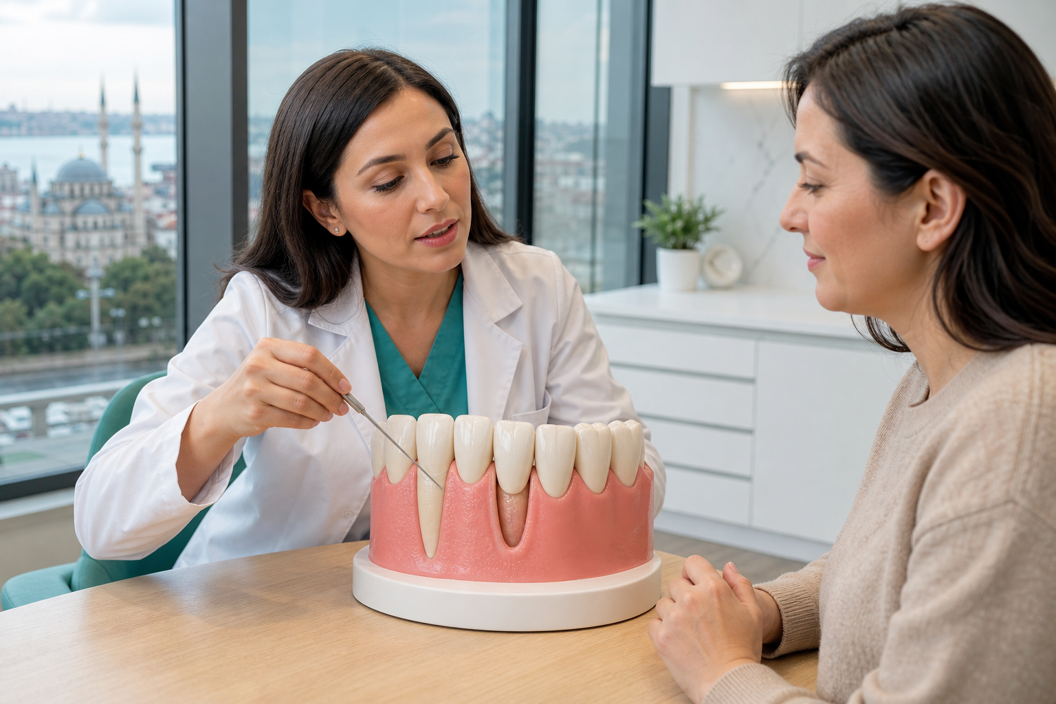
Clinical review required: Gum recession is the movement of the gum margin away from its previous position, exposing root surface. It may be associated with inflammation, thin tissues, tooth position, brushing trauma or periodontal attachment loss. Rapid change, pus, marked mobility, swelling or severe pain needs prompt assessment.
What are receding gums?
Gingival recession exposes part of a tooth root and makes a tooth appear longer. It can affect one tooth or many. Recession is a clinical finding rather than a diagnosis: the dentist must determine whether it reflects local anatomy, traumatic forces, periodontal disease, orthodontic movement, restoration contours or several factors together.
Common signs
People may notice a longer tooth, a notch near the gumline, root sensitivity, a darker or yellower root colour, food trapping or an uneven gum line. Some recession is painless and progresses slowly. Bleeding suggests inflammation but recession itself does not always bleed. Photographs and measurements over time help distinguish a stable defect from active change.
Why gums recede
Potential contributors include thin gum and bone phenotype, a tooth positioned toward the outside of the bony housing, plaque-induced inflammation, periodontitis, forceful brushing, traumatic oral habits, frenum pull, piercings and poorly contoured restorations. Smoking can mask bleeding while disease progresses. A single cause should not be assumed from appearance.
Recession versus periodontitis
Recession may occur without deep periodontal pockets or progressive bone loss. Periodontitis involves inflammatory destruction of the tooth-supporting attachment and bone. Both can coexist. Full periodontal charting, radiographs and risk assessment are therefore more informative than measuring the visible root alone.
Brushing and oral hygiene
Effective cleaning remains essential even when roots are exposed. Scrubbing aggressively with a hard brush can traumatise tissue and abrade root surface, but avoiding the area allows plaque inflammation to worsen. A clinician or hygienist can demonstrate a gentle technique, suitable brush and interdental method tailored to the defect.
Tooth sensitivity
Exposed dentine can react to cold, touch, sweet or acidic foods. Desensitising toothpaste, topical fluoride, varnish, bonding agents or a small restoration may reduce symptoms. Sensitivity can also come from decay, cracks or pulpal disease, so persistent or spontaneous pain should not be self-diagnosed as recession.
How recession is assessed
The examination records recession depth and width, probing depth, clinical attachment, bleeding, tissue thickness, keratinised tissue, root shape, cervical wear and tooth mobility. The dentist evaluates tooth position, bite, restorations and frenum. Radiographs show interproximal bone but do not map the thin facial bone reliably; CBCT is not routine for every recession defect.
Can gums grow back?
Lost gum margin does not usually regenerate through toothpaste, mouthwash or massage. Inflammation can settle and swelling can change, making the margin look different. Stable recession may need only prevention and monitoring. Surgical root-coverage procedures can move and thicken tissue in selected cases, but complete coverage is not guaranteed.
When monitoring is appropriate
A shallow, stable, cleansable recession without significant sensitivity, decay or aesthetic concern may be documented and reviewed. Monitoring includes standardised measurements or photographs and control of modifiable causes. Treatment is not automatically required because a root is visible.
Non-surgical management
First-line care may include plaque control, professional cleaning, desensitising products, fluoride, correction of traumatic brushing, removal of local irritants and management of active periodontal disease. A cervical composite restoration may protect a worn or decayed root but does not move the gum. Its contour must preserve cleaning access.
Orthodontic considerations
Moving a tooth toward a more favourable position within the alveolar housing may improve the environment for soft tissue, while movement outside the bony envelope may increase risk in susceptible anatomy. Orthodontic treatment does not reliably regenerate lost gum. Periodontal and orthodontic planning should agree on sequencing and tissue monitoring.
Root-coverage surgery
Common approaches include a coronally advanced flap or tunnel combined with a connective-tissue graft. A graft may be harvested from the palate; collagen matrices and other substitutes can avoid a donor site but may have different outcomes. Technique selection depends on the number and type of recessions, papilla and interproximal support, tissue phenotype and anatomy.
What affects surgical predictability?
Complete coverage is more predictable when interproximal attachment is preserved and the tooth is favourably positioned. Deep or wide defects, interproximal tissue loss, root prominence, cervical restorations, smoking, poor plaque control and unstable disease can limit coverage. Success may mean thicker, more maintainable tissue or reduced sensitivity rather than a perfectly hidden root.
Connective-tissue graft recovery
Swelling, tenderness and minor bleeding can occur at the recipient and donor sites. Patients receive site-specific cleaning, diet and medication instructions. Pulling the lip to inspect the graft can disturb healing. Increasing bleeding, fever, worsening swelling, graft displacement or uncontrolled pain should be reported promptly.
Recession around crowns and veneers
Recession may expose a restoration margin or darker root. Replacing the restoration can improve contour or colour but does not correct thin tissue or active inflammation. Periodontal stabilisation and an assessment of margin position should come before cosmetic replacement. Placing a new margin deeper under the gum may create another hygiene problem.
Recession around implants
Soft-tissue recession around an implant can reveal metal or restorative components. Causes and treatment differ from tooth recession because there is no periodontal ligament and implant surface decontamination may be relevant. The clinician checks implant position, bone, tissue volume, prosthesis contour and inflammation before considering grafting.
Preventing progression
Use a gentle effective brushing technique, clean interdental spaces, attend periodontal maintenance and report new sensitivity or visible change. Avoid oral jewellery that contacts the gum. Control smoking and active inflammation. A night guard may protect teeth from some load-related damage but is not a direct treatment for recession.
Treatment abroad and records
Request a full periodontal chart, recession classification, photographs, radiographs, tooth-position assessment, graft material and donor-site plan. Ask what amount of root coverage is realistic and how healing will be reviewed after travel. Clarify who manages donor-site bleeding, graft exposure or incomplete coverage.
Questions to ask
- Is the recession stable or progressing?
- Is periodontitis or interproximal bone loss present?
- Which causes can be modified?
- Is treatment for sensitivity, health, aesthetics or tissue thickness?
- What outcome is realistic with surgery?
- What maintenance protects the result?
Frequently asked questions
Does hard brushing cause every recession?
No. Brushing trauma can contribute, but anatomy, inflammation, tooth position and restorations also matter.
Can mouthwash rebuild gum?
No. It may support plaque control in selected cases but does not regenerate a lost gum margin.
Is grafting always necessary?
No. Stable, comfortable and cleansable defects may be monitored.
Will a graft last forever?
Long-term stability depends on defect anatomy, healing, hygiene, inflammation control and ongoing mechanical factors.
Sources and clinical review references
- Connective-tissue graft and coronally advanced flap: network meta-analysis.
- Root-coverage procedures with enamel matrix derivative: meta-analysis.
- Connective-tissue graft substitutes for gingival recession.
- Tunnel and coronally advanced flap techniques: systematic review.
Editorial review note: Evidence reviewed 22 July 2026. Named clinician review is required before indexation.