
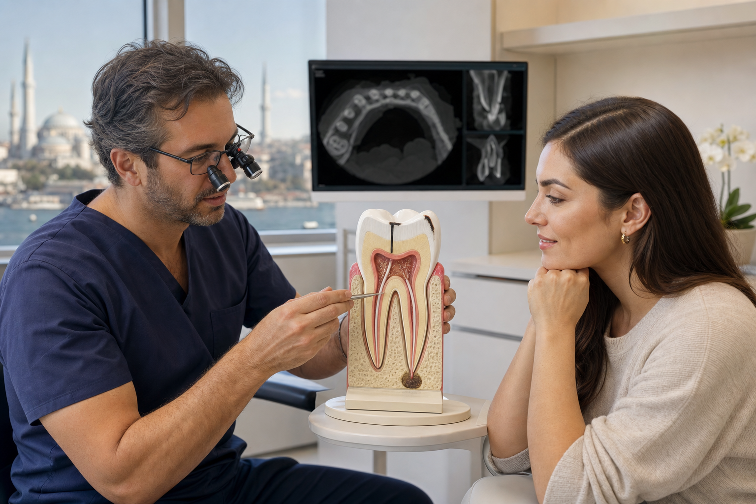
Clinical review required: Persistent symptoms or radiographic disease after root-canal treatment may arise from healing time, reinfection, missed anatomy, leakage, procedural complications, periodontal disease or root fracture. “Failure” requires diagnosis, not an X-ray label alone.
What is a failed root canal?
A root-treated tooth is considered unsuccessful when symptoms, infection or progressive apical disease persist or recur and the tooth requires further intervention. Healing is not immediate, and a radiographic lesion can take months or years to reduce. A tooth that is comfortable but not yet fully healed is not automatically a failure.
Symptoms
Possible signs include biting pain, spontaneous ache, swelling, a gum pimple, bad taste, mobility or repeated tenderness. Some persistent apical disease is asymptomatic and found on review imaging. Symptoms can also come from a crack, high bite, adjacent tooth, sinus or jaw muscles rather than the root canal.
Why treatment can fail
Causes include untreated canals, complex anatomy, residual infection, inadequate disinfection, short or poorly compacted filling, coronal leakage, new decay, a loose restoration, perforation, separated instruments and vertical root fracture. External infection or cyst-like lesions occasionally contribute. More than one cause may exist.
Missed canals
Molars commonly have variable canal anatomy. A missed canal can retain infected tissue and is associated with post-treatment apical periodontitis. Magnification, angled radiographs and selected CBCT can identify anatomy, but scans have artefacts and should be justified by how the result changes treatment.
Coronal leakage
A root filling needs a durable seal above it. Lost temporary fillings, recurrent decay, open crown margins and delayed final restoration allow contamination. Retreatment of canals without correcting the coronal pathway invites recurrence. Restorability and final restoration are assessed before endodontic procedures.
Vertical root fracture
A fracture can mimic endodontic failure with an isolated deep pocket, recurrent swelling or a J-shaped bone lesion. It may be difficult to prove. Removing the crown or filling, microscopy, probing and CBCT can help, but not every fracture is visible. A confirmed vertical root fracture often has a poor prognosis.
How healing is assessed
The clinician compares symptoms, examination and radiographs over time. Images should use similar angles where possible. Bone density can change slowly after adequate treatment. Enlarging lesions, new symptoms or persistent drainage carry different weight from a stable scar-like radiolucency.
CBCT
Three-dimensional imaging can reveal missed roots, untreated anatomy, resorption, perforation and cortical bone changes not clear on conventional radiographs. Metal and root-filling materials create artefact, and very fine fractures can remain invisible. CBCT is not a mandatory scan for every root-treated tooth.
Nonsurgical retreatment
Retreatment reopens the tooth, removes previous filling material, locates anatomy, disinfects and reseals canals. Existing crowns, posts, separated instruments and ledges can complicate access. The clinician balances the chance of correcting the cause against removal of additional tooth structure.
When retreatment is preferred
Orthograde retreatment is often considered when inadequate filling, missed anatomy or coronal leakage can be addressed from above and the tooth is restorable. It can treat the whole canal system rather than only the root end. A sound crown may need to be accessed or removed.
Apical surgery
Apicoectomy removes the root tip and inflamed tissue, then seals the canal from the root end. Modern microsurgery can be effective when orthograde access is impractical or previous retreatment has not resolved disease. It cannot correct an unrestorable crown or a vertical root fracture.
Root resection
In selected multirooted teeth, a diseased or fractured root may be removed while the remaining tooth is restored. Evidence is limited and maintenance is demanding. Root anatomy, periodontal support, furcation design and restorative plan determine whether it is realistic.
Extraction
Extraction is appropriate when the tooth is split, non-restorable, severely unsupported, repeatedly infected with poor correction prospects or strategically unsuitable. Replacement options include implant, bridge, denture, orthodontic closure or monitoring. An implant is not automatically simpler or complication-free.
Antibiotics
Antibiotics do not correct a missed canal, leaking crown or fracture. They are used for spreading infection, systemic signs or individual medical risk, alongside source control. Repeated prescriptions can suppress symptoms temporarily and contribute to adverse effects and resistance.
Pain after recent treatment
Mild biting tenderness for several days can be expected after treatment. Severe, increasing pain, swelling or a high bite requires review. Persistent symptoms do not prove infection; occlusion, crack, periodontal tissues and adjacent teeth should be reassessed before repeating treatment.
Restorability
Before retreatment, the dentist evaluates remaining tooth structure, cracks, ferrule, decay, periodontal support and the feasibility of a final restoration. Technically perfect retreatment has limited value if the tooth cannot be sealed or protected. Crown lengthening or orthodontic extrusion may occasionally improve restorability.
Success and prognosis
Outcome depends on the original cause, ability to disinfect and seal, lesion size, anatomy, fracture status, restoration and health. Population success rates cannot guarantee an individual tooth. The plan should include alternative pathways if healing does not occur.
Follow-up
Clinical and radiographic review is part of treatment. Report swelling, drainage, mobility or lost restoration promptly. A tooth can be comfortable while bone healing continues. Repeated CBCT solely to seek faster radiographic change is not justified without a clinical question.
Treatment abroad
Request original and postoperative radiographs, canal count, working-length and procedural records, material details and crown status. Ask whether a microscope and CBCT were used for a defined reason. Confirm who manages flare-up, perforation, separated instruments or persistent disease after travel.
Questions to ask
- Is this true persistent disease or expected healing?
- What is the most likely correctable cause?
- Is a fracture excluded?
- Can the tooth receive a durable final restoration?
- Why choose retreatment, surgery or extraction?
- How and when will healing be reviewed?
Frequently asked questions
Can a root canal fail years later?
Yes. New leakage, decay or fracture can reintroduce disease after a previously stable period.
Does a dark area mean failure?
Not by itself. Timing, symptoms and comparison with earlier images are essential.
Can the treatment be repeated?
Often, when a correctable cause exists and sufficient tooth structure remains.
Is extraction safer?
Not automatically. It removes the source but introduces replacement decisions and their own risks.
Sources and clinical review references
- Missed canals and post-treatment apical periodontitis.
- Nonsurgical retreatment versus apical surgery.
- Periapical healing after surgical endodontic retreatment.
- Root resection, retreatment and apical surgery outcomes.
Editorial review note: Evidence reviewed 22 July 2026. Named clinician review is required before indexation.